Highlights
The external eye has both anatomical and immunologic defense mechanisms for protection against infection and other ocular conditions.
Knowledge of corneal anatomy is vital for an understanding of corneal disease clas sifications and mastery of evolving keratoplasty techniques.
The cornea is a transparent and avascular tissue. Transparency results from the organization of keratocytes, fibers, and the extracellular matrix within the corneal stroma as well as the delicate balance of forces controlling stromal water content.
Eyelids
The functions of the eyelid include eye protection, tear distribution, ocular surface cleaning, regulation of light exposure, and contribution to the tear film. Except for fine vellus hairs, the eyelashes (cilia) are the only hairs of the eyelid. Eyelashes catch small particles and work as sensors to stimulate reflex eyelid closure. Blinking stimulates the lacrimal pump to release tears, which are then spread across the cornea, flushing foreign material. Most individuals blink an average of 10–15 times per minute at rest, 20 times per minute or more during a conversation, and as few as 5 times per minute when concentrating (eg, reading). Blink frequency also changes in different positions of gaze. The orbicularis oculi muscle, which is innervated by cranial nerve (CN) VII, closes the upper and lower eyelids (Fig 1-1). The leva tor palpebrae muscle, innervated by CN III, inserts into the tarsal plate and skin and elevates the upper eyelid. The Müller muscle, innervated by sympathetic nerves, also contributes to the elevation of the upper eyelid. The inferior tarsal muscle helps retract the lower eyelid.
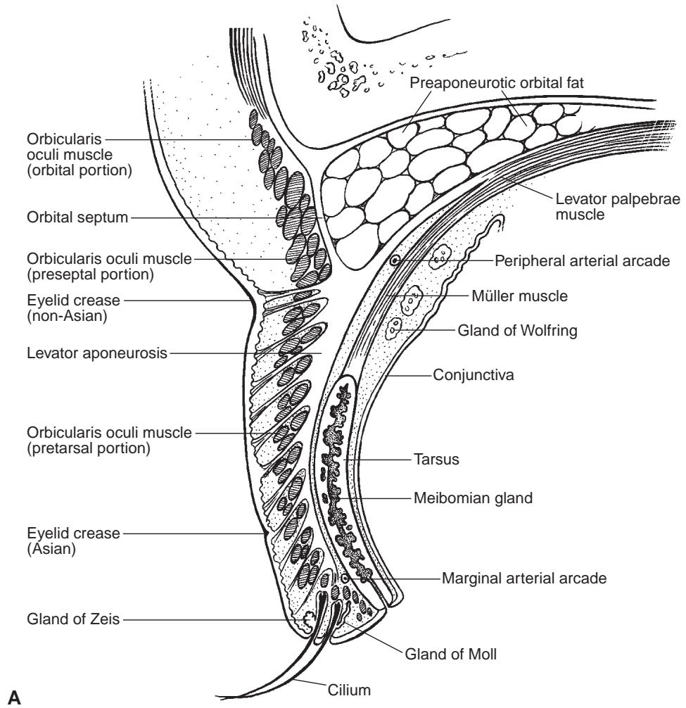
Figure 1-1 The eyelid. A, Illustration of a cross section of the upper eyelid.
(Continued)
The epidermis of the eyelids abruptly changes from keratinized to nonkeratinized stratified squamous epithelium at the mucocutaneous junction of the eyelid margin, along the row of meibomian gland orifices. Holocrine sebaceous glands and eccrine sweat glands are present in the eyelid skin. Near the eyelid margin are apocrine sweat glands (the glands of Moll) and numerous modified sebaceous glands (the glands of Zeis).
For additional discussion of eyelid anatomy, see BCSC Section 2, Fundamentals and Principles of Ophthalmology, and Section 7, Oculofacial Plastic and Orbital Surgery.
Argilés M, Cardona G, Pérez-Cabré E, Rodríguez M. Blink rate and incomplete blinks in six different controlled hard-copy and electronic reading conditions. Invest Ophthalmol Vis Sci. 2015;56(11):6679–6685.
Lin LK, Gokoffski KK. Eyelids and the corneal surface. In: Mannis MJ, Holland EJ, eds. Cornea. Vol 1. 4th ed. Elsevier; 2017:40–45.
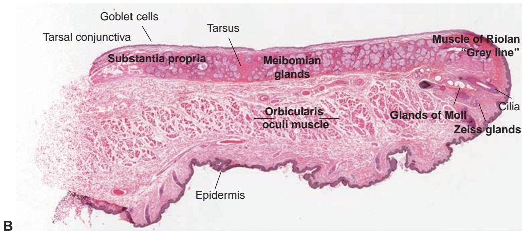
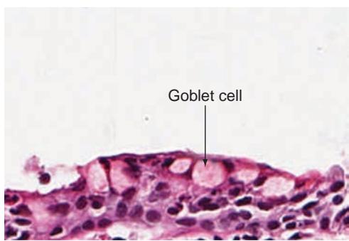
Figure 1-1 (continued) B, Hematoxylin-eosin (H&E) stained section of the normal eyelid. (Part A illustration by Christine Gralapp, part B © American Academy of Ophthalmology 2020.)
Lacrimal Functional Unit
The lacrimal functional unit (LFU) comprises the lacrimal glands, ocular surface, and eyelids, as well as the sensory and motor nerves that connect these components (Video 1-1; Fig 1-2). The LFU is responsible for the following:
regulation, production, health, and integrity of the tear film (carrying out lubricat ing, antimicrobial, and nutritional roles)
health of the ocular surface (maintaining corneal transparency and the surface stem cell population)
quality of the image projected onto the retina

VIDEO 1-1 Perception of touch and innervation of the lacrimal functional unit.
Modified with permission from Pflugfelder SC, Beuerman RW, Stern ME, eds. Dry Eye and Ocular Surface Disorders. Published by CRC Press. © Marcel Dekker; 2004, reproduced by arrangement with Taylor & Francis Books UK.

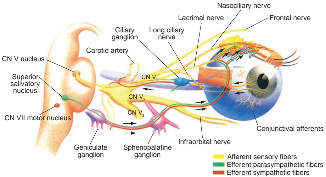
Figure 1-2 The sensory and motor nerves connecting the components of the lacrimal func tional unit. CN = cranial nerve. (Modified with permission from Pflugfelder SC, Beuerman RW, Stern ME, eds. Dry Eye and Ocular Surface Disorders. Published by CRC Press. @ Marcel Dekker; 2004, reproduced by arrangement with Taylor & Francis Books UK.)
The LFU responds to environmental, endocrinologic, and cortical influences. The afferent component of the LFU is mediated through ocular surface and trigeminal no ciceptors, which synapse with the efferent nerves (autonomic and motor nerves) in the brainstem. The autonomic nerve fibers innervate the meibomian glands, conjunctival gob let cells, and lacrimal glands. The motor nerve fibers innervate the orbicularis muscle to initiate blinking. During blinking, the meibomian glands express lipid, and the tears are replenished from the inferior tear meniscus and spread across the cornea while excess tears are directed into the lacrimal puncta.