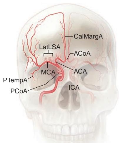
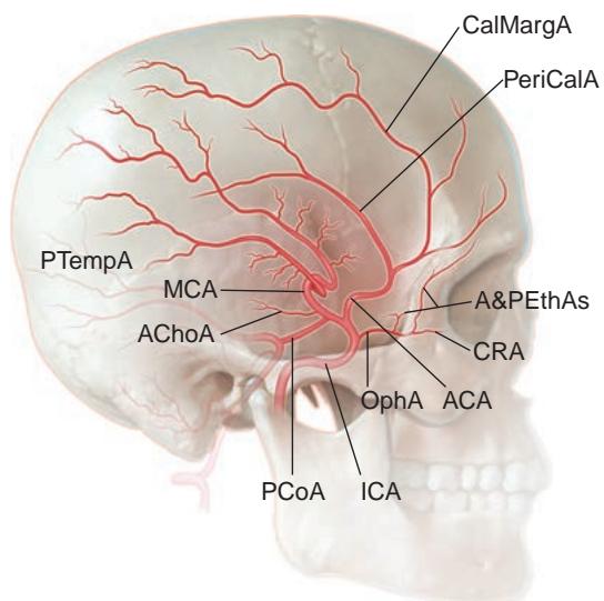
Figure 1-10 Branches of the internal carotid artery (ICA). A, Lateral view. B, Anteroposterior view. Proximal ICA branches: ophthalmic artery (OphA) and its branches (anterior and poste rior ethmoidal arteries [A&PEthAs] and central retinal artery [CRA]); posterior communicating artery (PCoA); anterior choroidal artery (AChoA); anterior communicating artery (ACoA); ante rior cerebral artery (ACA) and its branches (callosomarginal artery [CalMargA] and pericallosal artery [PeriCalA]); and middle cerebral artery (MCA) and its branches (lateral lenticulostriate artery [LatLSA] and posterior temporal artery [PTempA]). (Illustrations by Rob Flewell, CMI.)
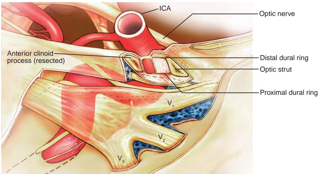
Figure 1-11 Lateral view of the bend of the internal carotid artery (ICA) within the right cavernous sinus. The anterior clinoid process has been resected to show the proximal and distal dural rings. $V _ { 1 } = $ ophthalmic division of CN V; $V _ { 2 } = $ maxillary division of CN V; $V _ { 3 } = $ mandibular division of CN V. (Illustration by Dave Peace.)
retinal arteries and arterioles have tight junctions that form a blood–retina barrier, much like the blood–brain barrier. The intraretinal arterioles run within the substance of the nerve fiber layer to supply the inner two-thirds of the retina.
CLINICAL PEARL
In most people, the OphA runs under the intracranial portion of the optic nerve. Therefore, an OphA aneurysm causes an inferior visual field defect because the superior portion of the optic nerve is pushed up against the falciform ligament.
The lacrimal artery, which also derives from the OphA, runs parallel to the lacrimal branch of CN V1 in the superior lateral orbital roof to reach the lacrimal gland. It also gives off the anterior ciliary artery of the lateral rectus muscle, which reaches the anterior segment at the muscle’s insertion. The frontal artery runs within the superior orbit, paral leling the frontal branch of CN V1 and separates into and terminates as the supraorbital and supratrochlear arteries that, along with the lacrimal artery, supply the eyelid.
The next branches leaving the OphA are the superior and inferior muscular arteries, which supply the anterior ciliary arteries of the superior rectus and superior oblique muscles (superior muscular branch) and the medial and inferior rectus muscles (inferior muscular artery). The anterior ciliary arteries are responsible for most of the blood flow to the cili ary body. The medial and lateral long posterior ciliary arteries sometimes anastomose with penetrating branches of the anterior ciliary arteries (within the rectus muscles) to form the greater arterial circle near the anterior part of the ciliary body. Branches from this circle extend radially within the iris to form a second anastomotic circle (the lesser arterial circle) near the collarette of the iris.
The terminal OphA supplies additional branches that form collaterals with the ante rior and posterior ethmoidal arteries and also form the short and long posterior ciliary ar teries (Fig 1-12). The short posterior ciliary arteries then divide into 10–20 small branches that supply the optic nerve head (ONH) and posterior choroid. Together, these arter ies also supply the retinal pigment epithelium and approximately the outer one-third of the retina, including the photoreceptor cells. In approximately 20%–30% of individuals, branches of the posterior ciliary arteries (cilioretinal arteries) directly supply a portion of the inner retina; this blood supply may protect the macula in the case of a CRA occlusion. Approximately 4 short posterior ciliary arteries form a variably complete anastomotic ring (known as the circle of Zinn-Haller) around the ONH, which is also supplied by the peri papillary choroid and the terminal branches of the pial network.
Collateral branches from terminal branches of the infraorbital artery and the superfi cial temporal artery help supply the lower and upper eyelids and may also provide collat eral supply to the anterior segment. These collaterals may be interrupted if the conjunctiva and Tenon capsule are removed from the limbus during ocular surgery.
Distal to the origin of the OphA, the intradural supraclinoid ICA gives off the anterior choroidal artery (AChoA) and anastomoses with the proximal PCA through the posterior communicating artery (PCoA). The AChoA supplies blood proximally to the optic tract and distally to the lateral geniculate nucleus (LGN) (Fig 1-13). Occlusion of the AChoA can produce optic tract syndrome, a disorder consisting of contralateral homonymous hemi anopia, contralateral band atrophy of the ONH, and a contralateral relative afferent pu pillary defect (RAPD; see Chapter 4). The ICA then gives off the anterior cerebral artery (ACA) and terminates as branches of the middle cerebral artery (MCA). The proximal ACA (the A1 segment) crosses over the optic nerve and joins the opposite ACA via the anterior communicating artery (ACoA). This combination of the ACoA and PCoA creates the circle of Willis, which permits collateral flow between the carotid and vertebrobasilar systems when there is vascular compromise (Fig 1-14). Small perforating branches arising from the proximal ACA as well as the ACoA also supply the intracranial optic nerves and chiasm (Fig 1-15).
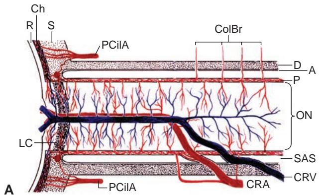
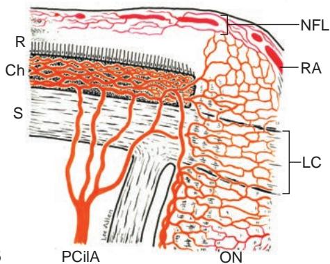
Figure 1-12 Schematic representation of the vascular supply to the optic nerve and optic nerve head (ONH). A, Lateral view of the ONH. B, Axial view of the ONH. Short posterior ciliary arteries supply the centripetal capillary beds of the anterior ONH. The central retinal artery (CRA) contri bution is restricted to capillaries of the nerve fiber layer and the anterior intraorbital optic nerve. Capillary beds at all levels drain into the central retinal vein (CRV). A = arachnoid; Ch = choroid; ColBr = collateral branch; D = dura; LC = lamina cribrosa; NFL = surface nerve fiber layer of the ONH; ON = optic nerve; P = pia; PCilA = posterior ciliary artery; R = retina; RA = retinal arteriole; S = sclera; SAS = subarachnoid space. (Reprinted from Hayreh SS. The blood supply of the optic nerve head and the evaluation of it—myth and reality. Prog Retin Eye Res. 2001;20(5):563–593, with permission from Elsevier.)