
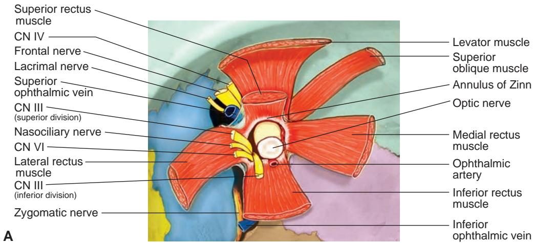
Figure 1-5 Anatomy of the orbital apex. A, The 4 rectus muscles arise from the annulus of Zinn. CNs II, III (superior and inferior branches), and VI and the nasociliary nerve all course through the annulus of Zinn. CN IV, the frontal and lacrimal nerves, and the ophthalmic veins are located outside the annulus. B, Anatomical dissection just anterior to the superior orbital fissure. (Part A illustration by Dave Peace; part B courtesy of Albert L. Rhoton Jr, MD.)
Under the apex of the orbit, the pterygomaxillary area contains the sphenopalatine ganglion and the internal maxillary artery. The pterygomaxillary area communicates
anteriorly through the infraorbital canal to the cheek and lower eyelid
posteriorly through the foramen rotundum and vidian canal to the middle cranial fossa
superiorly through the inferior orbital fissure to the orbit
B

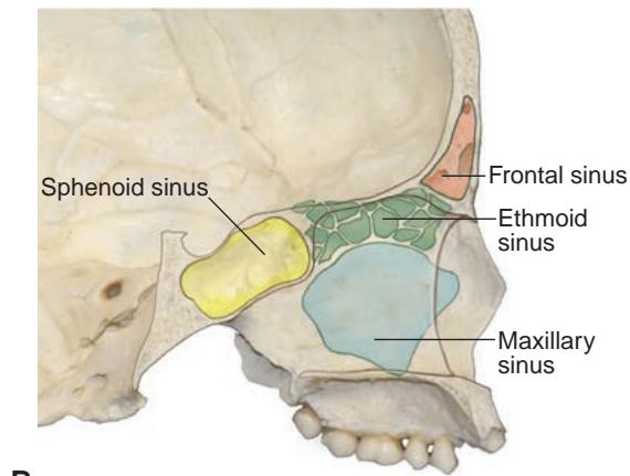
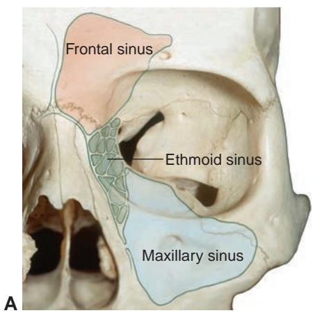
Figure 1-6 Anatomical relationship of the 4 paranasal sinuses to the orbit. A, Coronal view. B, Sagittal view. C, Axial view. (Illustrations by Dave Peace.)
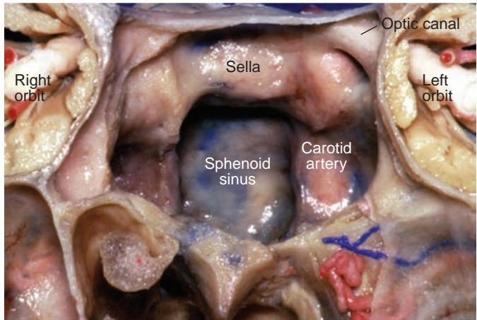
Figure 1-7 Coronal section, anterior view into the sphenoid sinus demonstrating the relation ship of the internal carotid artery and optic nerve within the lateral wall of the sinus. (Courtesy of Albert L. Rhoton Jr, MD.)
Canals and fissures
The orbit communicates with the surrounding areas through several bony canals and fis sures. Posteriorly, the orbit is contiguous with the cavernous sinus through the superior orbital fissure (see Fig 1-3). The medial wall of the orbit continues as the lateral wall of the sphenoid bone, marking the medial extent of the cavernous sinus. When sharp objects enter the me dial orbit, they are directed through the superior orbital fissure, where they can lacerate the carotid artery. Within the cavernous sinus, iatrogenic or traumatic damage to the internal ca rotid artery (ICA) may produce a direct, high-flow carotid-cavernous fistula (see Chapter 8).
The orbit is connected superiorly and posteriorly to the anterior cranial fossa by way of the optic canal (see Fig 1-2), which transmits the optic nerve, OphA, and some of the oculo sympathetic fibers (terminal branches of the carotid plexus). Inferiorly at the apex, the orbit is connected to the pterygopalatine fossa—and thus the temporal and inferotemporal regions— through the inferior orbital fissure. This fissure, formed by the greater wing of the sphenoid bone and the maxillary, zygomatic, and palatine bones, carries the following structures:
maxillary branch of CN V (CN V2)
infraorbital vessels
inferior ophthalmic vein
branches from the pterygopalatine ganglion
parasympathetic fibers that innervate the lacrimal gland
collateral meningeal arteries that connect the external and internal carotid circulation
Anteriorly, the orbit connects to the inferior meatus of the nose (beneath the inferior turbinate) through the nasolacrimal duct, which carries tears into the nasal cavity. Other canals surrounding the orbit include the anterior and posterior ethmoidal foramina, which carry blood vessels that connect the internal carotid circulation (ie, the OphA) to the ex ternal carotid circulation (ie, the terminal branches of the ethmoidal arteries). Additional supraorbital and zygomaticotemporal foramina carry blood vessels between the orbit and corresponding vessels of the forehead and temple.
Rhoton AL, Natori Y. The Orbit and Sellar Region: Microsurgical Anatomy and Operative Approaches. Thieme; 1996.