The parasellar region is connected laterally to the petrous and temporal bones and inferiorly to the clivus, extending to the foramen magnum and the exit of the spinal cord. The posterior skull base is enclosed by the occipital bones (Fig 1-3).
The Orbit
The anterior aspect of the skull includes the orbits, which connect posteriorly to the para sellar region. Approximately 45 mm wide and 35 mm in maximal height, the orbit has a total volume of about 30 cm3. Its medial walls are about 40 mm from the rim to the optic canal. Although these walls are roughly parallel, the lateral walls form an almost 90° angle.
The orbit is composed of 7 craniofacial bones (Fig 1-4, Table 1-1):
maxillary
zygomatic
frontal
lacrimal
sphenoid
palatine
ethmoid
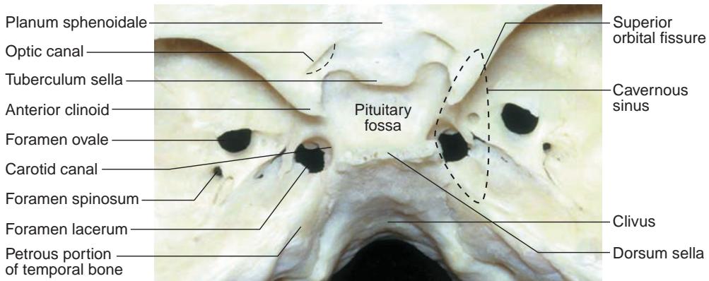
Figure 1-3 Parasellar bony anatomy demonstrates the relationship of the pituitary fossa to the cavernous sinus, including the foramina of the skull base. The foramen lacerum is filled with cartilage and contains the artery of the pterygoid canal, the nerve of the pterygoid canal, and the venous drainage structures. The carotid artery enters the skull base through the carotid canal. (Courtesy of Albert L. Rhoton Jr, MD.)
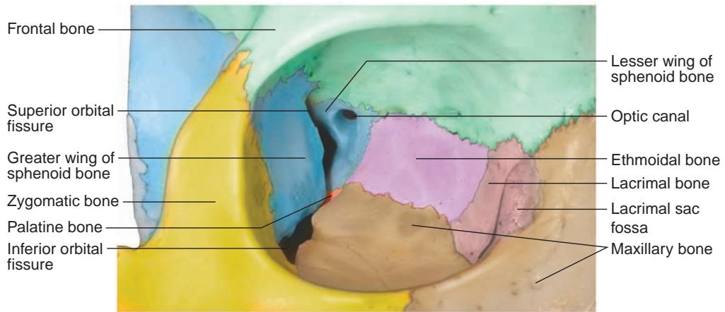
Figure 1-4 Anatomy of the orbit. Bony anatomy of the right orbital apex. The optic canal trans mits the optic nerve, ophthalmic artery, and some oculosympathetic fibers. The superior orbital fissure, between the greater and lesser wings of the sphenoid bone, transmits CNs III, IV, and VI; the ophthalmic division of CN V (CN V1); the oculosympathetics; and the superior ophthalmic vein. (Illustration by Dave Peace.)
Table 1-1 Bones of the Orbit
| Orbital Roof | Lateral Wall | Orbital Floor | Medial Wall |
| Frontal | Zygomatic | Zygomatic | Maxillary |
| Lesser wing of sphenoid | Greater wing of sphenoid | Maxillary | Lacrimal |
| Palatine | Ethmoid | ||
| Lesser wing of sphenoid |
The superior orbital rim is made up of the frontal bone, which connects to the zygomatic bone laterally at the frontozygomatic suture. The inferior orbital rim is made up of the zygomatic bone inferolaterally and the maxillary bone inferonasally, which meet at the zy gomaticomaxillary suture. Medially, the orbital rim consists of the maxillary and lacrimal bones, which join the frontal bone superiorly. Three additional bones contribute to the orbit: the ethmoid bone medially, the palatine bone inferiorly in the posterior orbit, and the sphenoid bone laterally and superiorly in the orbital apex (Activity 1-1).

ACTIVITY 1-1 Bony anatomy of the orbit. Developed by Zoë R. Williams, MD. Illustrations by Dave Peace.
At the orbital apex, the annulus of Zinn gives rise to the 4 rectus muscles. CNs II and III, the nasociliary nerve of CN V, and CN VI pass through the annulus of Zinn. In contrast, CN IV and the frontal and lacrimal nerves of CN V, as well as the superior ophthalmic vein, pass through the superior orbital fissure outside the annulus of Zinn (Fig 1-5).
CLINICAL PEARL
Because the superior oblique muscle is innervated by CN IV, which bypasses the annulus of Zinn, it is often not paralyzed—or is the last extraocular muscle to be paralyzed—by a retrobulbar block.
The orbit is surrounded by several important structures, including 4 paranasal sinuses (Fig 1-6):
the maxillary sinus, which is adjacent to the orbital floor
the ethmoid sinus, which is adjacent to the orbital medial wall
the sphenoid sinus, also adjacent to the orbital medial wall
the frontal sinus, with variable relationship to the anterior orbital roof
The sphenoid sinus forms the medial wall of the optic canal (Fig 1-7). In approxi mately 4% of patients, the bone may be incomplete, leaving only mucosa separating the sinus from the optic nerve. Surgery within the sphenoid sinus can potentially damage the optic nerve. In patients with pituitary or suprasellar lesions, use of the sphenoid sinus for an endoscopic surgical approach facilitates decompression of the optic chiasm. However, decompression can also be approached via craniotomy depending on the extent and site of pathology.
Other major structures around the orbit are the anterior cranial fossa superiorly (containing the frontal lobe) and the temporal fossa laterally (containing the temporalis muscle). The roof of the ethmoidal complex, delineated by the frontoethmoidal suture (top of the ethmoid bone, or lamina papyracea), marks the inferior boundary of the anterior cranial fossa. Surgical intervention above the frontoethmoidal suture, as occurs during an endoscopic sinus procedure, can result in inadvertent entry into the anterior cranial fossa or a cerebrospinal fluid (CSF) leak.