Objectives
Upon completion of BCSC Section 5, Neuro-Ophthalmology, the reader should be able to
explain a symptom-driven approach to the assessment of patients with common neuro-ophthalmic clinical manifestations and state an appropriate differential diagnosis
select the most appropriate diagnostic tests and imaging studies, based on clinical signs and symptoms
develop a management algorithm for neuro-ophthalmic disorders
identify neuro-ophthalmic emergencies
describe a plan for the immediate management of patients with neuro-ophthalmic emergencies
identify the anatomical structures that are relevant to neuroophthalmic disorders (including the skull base, orbit, brain, vascular system, and cranial nerves) in order to localize lesions
describe eye movement disorders and their anatomical relationship to the ocular motor system
describe the management of diplopia
state the various clinical manifestations and underlying causes of illusions, hallucinations, and cortical visual impairment
describe pupil, eyelid position, and ocular motor pathology in relation to specific disease processes
explain functional neurological symptom (conversion) disorder, factitious disorder, and malingering, and list the various clinical tests that are used to evaluate functional vision loss
describe the neuro-ophthalmic effects of the most common neurologic and systemic disorders affecting the visual and ocular motor systems
Neuro-Ophthalmic Anatomy

This chapter includes a related activity. Go to www.aao.org/bcscactivity_section05 or scan the QR code in the text to access this content.
Highlights
The optic canal is located within the lesser wing of the sphenoid bone and transmits the optic nerve, ophthalmic artery, and some oculosympathetic fibers.
The ophthalmic artery gives off the central retinal artery, which enters the optic nerve 10–12 mm posterior to the globe.
The optic chiasm is anterior to the hypothalamus and the anterior third ventricle and is usually approximately 10 mm above the sella.
Cranial nerve (CN) IV is the only CN that exits on the dorsal surface of the brain stem and has the longest unprotected intracranial course (likely responsible for its frequent involvement in closed-head trauma).
A central or upper motor neuron CN VII palsy causes contralateral lower facial weakness, whereas a peripheral or lower motor neuron CN VII palsy causes ipsilat eral upper and lower facial weakness.
Introduction
Although an adequate understanding of physiology—and increasingly molecular genetics— is important in managing diseases and potential treatments, anatomy remains the founda tion of medical practice in general and of surgical subspecialties in particular. Nowhere is this more apparent than in ophthalmology, given the complex structure and components of the human eye. This chapter outlines the intracranial pathways subserving the afferent and efferent visual pathways, the sensory and motor anatomy of the face, and the auto nomic nervous system of the eye and visual system. The anatomy of the globe and adnexal structures is covered in more detail in BCSC Section 2, Fundamentals and Principles of Ophthalmology; Section 7, Oculofacial Plastic and Orbital Surgery; and Section 8, External Disease and Cornea.
Bony Anatomy
Skull Base
The skull base has an intimate relationship with the visual system. It is connected to the lower facial skeleton by 3 sets of pillars formed by the maxillary and zygomatic bones an teriorly and the pterygoid process of the sphenoid bone posteriorly. Superiorly, the vault of the skull is made up of the following:
the parietal bones, which meet at the sagittal suture
the frontal bone, which adjoins the parietal bones at the coronal suture
the occipital bone, which meets the parietal bones at the lambdoid suture
See BCSC Section 6, Pediatric Ophthalmology and Strabismus, for more on these skull sutures.
In the skull base, located posterior and medial to the 2 orbits, is the sella turcica, a de pression within the body of the sphenoid bone (Fig 1-1). The lesser wing of the sphenoid bone is pierced by the optic canal, which allows the optic nerves to exit from the orbits. Within the canal, the optic nerve is accompanied by the ophthalmic artery (OphA) inferi orly and is separated from the superior orbital fissure by the optic strut (the posterior root of the lesser wing of the sphenoid), which terminates superiorly as the anterior clinoid (Fig 1-2). As a result, anterior clinoid meningiomas may cause compressive optic neu ropathy. Medially, the optic nerve is separated from the sphenoid sinus by bone that may be thin or dehiscent.
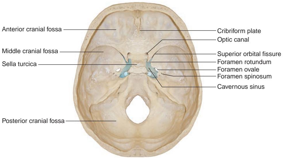
Figure 1-1 Bony anatomy of the skull base. The cavernous sinuses are located on each side of the sella turcica. Important openings within the skull base include the cribriform plate (transmits branches of the olfactory nerve, also known as cranial nerve [CN] I), optic canal (transmits the optic nerve, CN II), foramen ovale (transmits the mandibular division $ \mathrm{C N~ V_{3}} $ )of the trigeminal nerve, CN V), foramen rotundum (transmits the maxillary division $ \mathrm{C N~ V_{2}} $ )of CN V), superior orbital fissure (transmits CNs III, IV, VI, and V [ophthalmic division, CN $ V_{1} $ ), and foramen spinosum (transmits the middle meningeal artery, a branch of the external carotid artery). (Courtesy of Albert L. Rhoton Jr, MD.)
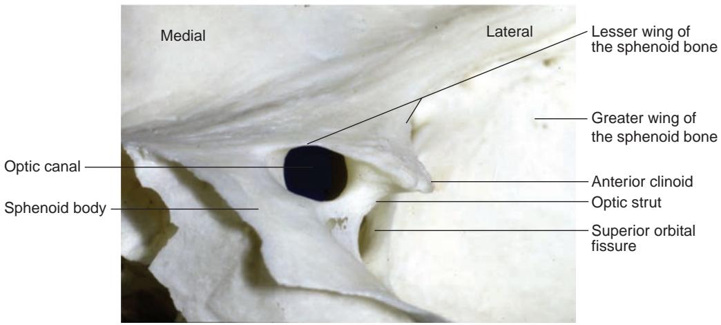
Figure 1-2 Intracranial view of the left optic canal. Within the lesser wing of the sphenoid bone is the optic foramen, which leads to the optic canal. The optic strut separates the optic canal from the superior orbital fissure. (Courtesy of Albert L. Rhoton Jr, MD.)
The gap between the lesser and greater wings of the sphenoid bone represents the superior orbital fissure, which transmits the following neurovascular structures:
oculomotor nerve (CN III)
trochlear nerve (CN IV)
ophthalmic division of the sensory trigeminal nerve (CN V1)
abducens nerve (CN VI)
oculosympathetic fibers
superior ophthalmic vein