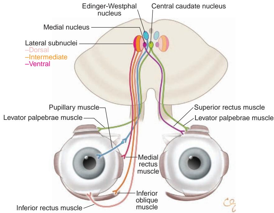
Figure 1-36 Oculomotor nucleus complex. Note that with the exception of the superior rectus muscle, all extraocular muscles served by CN III are innervated by their respective ipsilateral nuclei. Parasympathetic fibers travel to the pupillary sphincter muscle synapse in the ciliary ganglion in the orbit (see Fig 1-47). (Illustration by Christine Gralapp.)
Within the midbrain, CN III is topographically organized into a superior division (supplying the superior rectus and levator palpebrae superioris muscles) and an inferior division (supplying the medial and inferior rectus, inferior oblique, pupillary sphincter, and ciliary body muscles). However, the true anatomical division into 2 branches occurs at the level of the anterior cavernous sinus/superior orbital fissure (discussed in a follow ing paragraph).
The fascicles of CN III traverse the ventral midbrain tegmentum, passing near and possibly through the red nucleus, the substantia nigra, and the corticospinal tracts within the cerebral peduncle. The fascicles exit on the ventral surface of the peduncles (see Fig 1-35). Although CN III is considered a single structure within the subarachnoid space, as is the case in the midbrain, the nerve and its various fibers are topographically organized. Within the subarachnoid space, the nerve passes between the SCA below and the PCA above. It runs slightly oblique to the tentorial edge, parallel and lateral to the PCoA. The pupillary fibers are usually found on the dorsomedial surface of the nerve, where they are anatomically vulnerable to compression. The uncus, which is the most me dial aspect of the temporal lobe, is located just above the tentorium and the subarachnoid portion of CN III. Unilateral supratentorial mass lesions may force the uncus through the tentorial notch (uncal herniation) to compress the ipsilateral CN III.
At the back edge of the dura of the clivus and cavernous sinus, the nerve enters its own dural canal just above CN IV (see Fig 1-35). Running forward in the superior lateral wall of the cavernous sinus, the nerve separates into 2 divisions: superior and inferior. These divisions enter the orbit through the superior orbital fissure within the annulus of Zinn. The superior division runs forward intraconally to innervate first the superior rec tus muscle and then the levator palpebrae superioris muscle. The inferior division sends parasympathetic fibers to the ciliary ganglion in the orbital apex approximately 10 mm anterior to the annulus of Zinn and lateral to the optic nerve. Within the ciliary ganglion, the fibers destined for the pupillary sphincter and the ciliary body synapse. The fibers subsequently accompany the branch destined for the inferior oblique muscle. The number of fibers associated with accommodation innervating the ciliary body is approximately 9 or 10 times the number of fibers reaching the pupillary sphincter muscle. This disparity may be one reason for the development of light–near dissociation in Adie pupil (see Chap ter 11). The remaining branches of CN III within the orbit innervate the medial rectus and inferior rectus muscles.
Fourth cranial nerve
The CN IV (trochlear) nucleus lies within the gray matter in the dorsal aspect of the cau dal midbrain just below the aqueduct, directly contiguous with the more rostral CN III nu cleus (Fig 1-37). The intra-axial portion (fascicle) of CN IV is very short, running dorsally around the periaqueductal gray to cross within the anterior medullary velum just caudal to the inferior colliculi and below the pineal gland. CN IV is the only CN that exits on the dorsal surface of the brainstem, and it has the longest unprotected intracranial course (which is likely responsible for its frequent involvement in closed-head trauma). Within the subarachnoid space, CN IV swings around the midbrain, paralleling the tentorium just under the tentorial edge. As a result, CN IV is easily damaged during neurosurgical procedures that involve the tentorium.
The fourth CN enters the posterior lateral aspect of the cavernous sinus below the anterior tentorial insertion and underneath CN III. Covered by a variable sheath, CN IV runs forward within the lateral wall of the cavernous sinus. Anteriorly, the nerve crosses over CN III to enter the superior orbital fissure. Along with the frontal and lacrimal nerves of CN V and the SOV, CN IV passes through the superior orbital fissure but outside the annulus of Zinn. CN IV subsequently crosses over the optic nerve to enter the superior oblique muscle within the superior medial orbit.
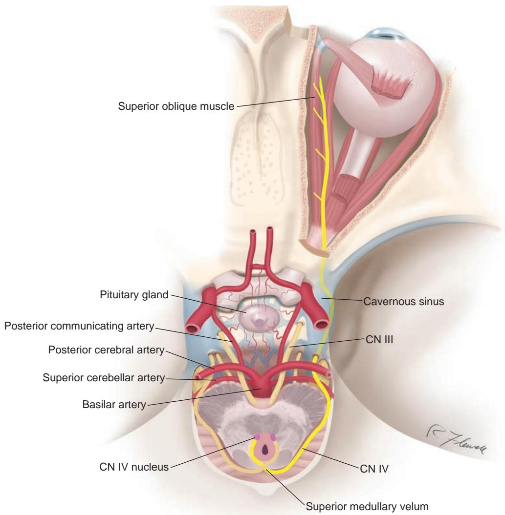
Figure 1-37 Illustration of the axial course of CN IV from its origin in the midbrain to the su perior oblique muscle. (Reprinted from Al-Zubidi N, Chevez-Barrios P, Foroozan R, Bhatti MT. My eyes are turned outside. Surv Ophthalmol. 2014;59(3):356, with permission from Elsevier.)
Sixth cranial nerve
Cranial nerve VI (abducens nerve) originates in the dorsal caudal pons just beneath the fourth ventricle. Its nucleus is surrounded by the looping fibers (genu) of CN VII and ad jacent to the PPRF and the MLF (Fig 1-38). The CN VI nucleus contains primary moto neurons and interneurons that cross to the contralateral MLF to reach the CN III nucleus. Pathology affecting the CN VI nucleus produces an ipsilateral gaze palsy.
The motor axons exiting the CN VI nucleus travel ventrally and slightly laterally, me dial to the superior olivary nucleus, emerging on the ventral surface of the caudal pons. As the fascicles pass through the brainstem, they lie adjacent to the spinal tract of CN V and traverse the corticobulbar tracts. Exiting the brainstem, the nerve runs rostrally within the subarachnoid space on the surface of the clivus from the area of the cerebel lopontine angle to the superior portion of the posterior fossa. The nerve pierces the dura approximately 1 cm below the petrous apex and travels beneath the petroclinoid liga ment (the Gruber ligament, which connects the petrous pyramid to the posterior clinoid) to enter the canal of Dorello. Within the canal, CN VI travels with the inferior petrosal sinus. Once it becomes extradural, the nerve is within the cavernous sinus, where it runs parallel to the horizontal segment of the carotid artery and CN V1 (Fig 1-39). CN VI is the only CN within the substance of the cavernous sinus; ipsilateral CN VI palsy and Horner syndrome localize here as the nerve is joined for a short segment by branches of the oculosympathetics lying within the wall of the intrapetrous carotid artery. Reaching the anterior portion of the cavernous sinus, CN VI traverses the superior orbital fissure through the annulus of Zinn (see Fig 1-5) to enter the medial surface of the lateral rectus muscle.
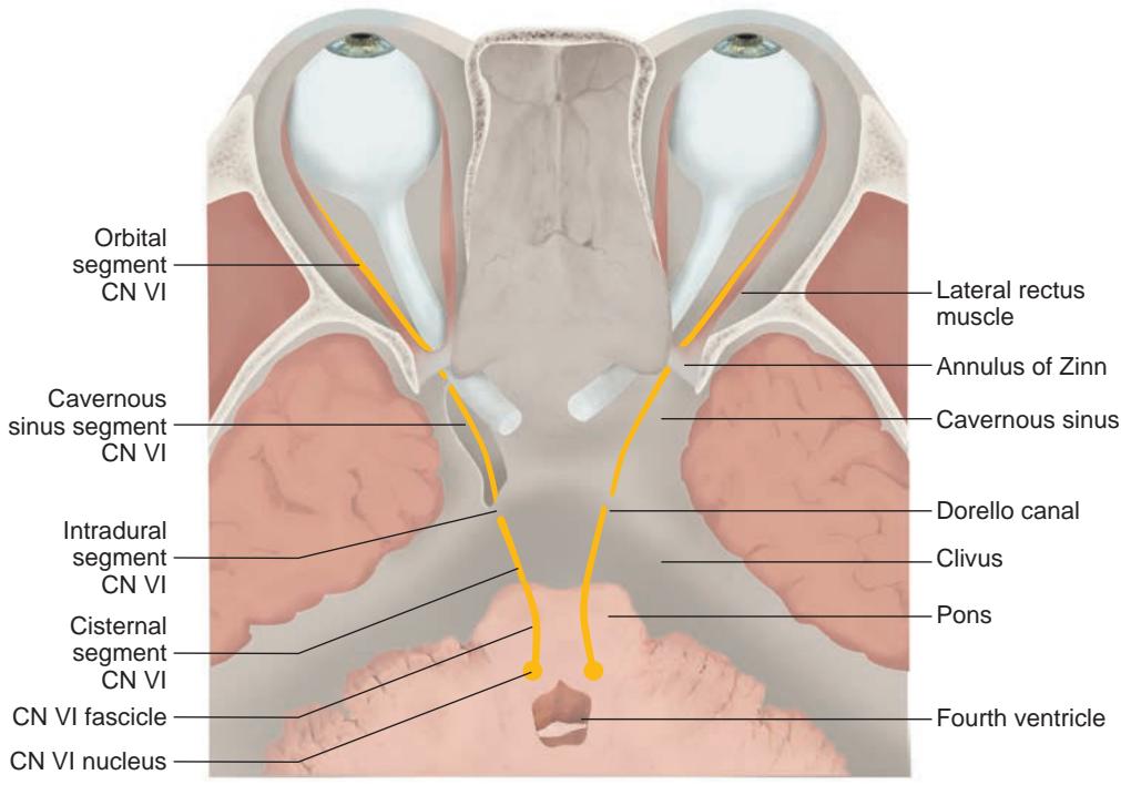
Figure 1-38 Illustration of the axial course of CN VI from its origin in the pons to the lateral rectus muscle. (Illustration by Cyndie C. H. Wooley.)