Smooth-pursuit system
The smooth-pursuit system consists of the following structures (Fig 1-30):
cortical structures: areas V1, V2, V3, V5 (middle temporal visual area), medial supe rior temporal area, posterior parietal cortex, and frontal eye field
subcortical structure: posterior portion of the internal capsule
brainstem neural network or premotor neurons: dorsolateral pontine nuclei and lat eral pontine nuclei
projections to the cerebellum (flocculus and dorsal vermis) and vestibular nuclei
For information on clinical disorders of the pursuit function, see Chapter 9 in this volume.
Subcortical Structures
Several subcortical structures are involved in relaying information between the cortex and the brainstem for coordination of eye movements:
SC: divided into 2 parts:
• superficial (dorsal): receives sensory input from the visual cortex and retina
• deep (ventral)—stratum griseum profundum and stratum album profundum: gen erate the motor signal to premotor structures
BG (caudate nucleus, putamen, and substantia nigra pars reticulata): controls volun tary saccades and inhibits unnecessary reflexive saccades
thalamus (internal medullary lamina and pulvinar): programs saccades

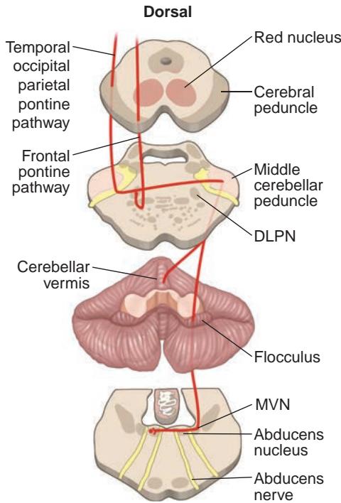
Figure 1-30 Pursuit system. A moving object creates signals in the striate cortex (V1) and from there to the extrastriate cortex (V2 and V3). These signals are subsequently relayed to the middle temporal (MT) and medial superior temporal (MST) visual areas, which are part of the dorsal visual processing stream, the pos terior parietal cortex, and the frontal eye field (FEF). The descending cortical pathways of the temporal, parietal, and frontal lobes inner vate the ipsilateral dorsolateral pontine nucleus (DLPN). The neurons in the DLPN decussate and project to the vermis and flocculus in the contralateral cerebellar lobe, which innervates the medial vestibular nucleus (MVN). VN neu rons then decussate and project to the con tralateral CN VI nucleus. The nucleus of CN VI initiates conjugate horizontal eye movements by innervating the ipsilateral lateral rectus mus cle and, via internuclear neurons that travel in the medial longitudinal fasciculus, the contra lateral medial rectus muscle. (Illustration by Rob Flewell, CMI.)
Brainstem
The following list of major anatomical structures and their functions lays the foundation for the discussion of pathways for coordinating conjugate eye movements in Chapter 9 (Fig 1-31):
riMLF: excitatory burst neurons that generate vertical and torsional saccades
interstitial nucleus of Cajal (INC): inhibitory burst neurons for vertical saccades and the neural integrator for vertical and torsional gaze
posterior commissure: projecting axons from the INC to the contralateral nuclei of CNs III, IV, and VI and the INC
medial longitudinal fasciculus (MLF): major pathways for relaying signals within the brainstem and for connecting the nuclei of CN VI to the nuclei of CN III
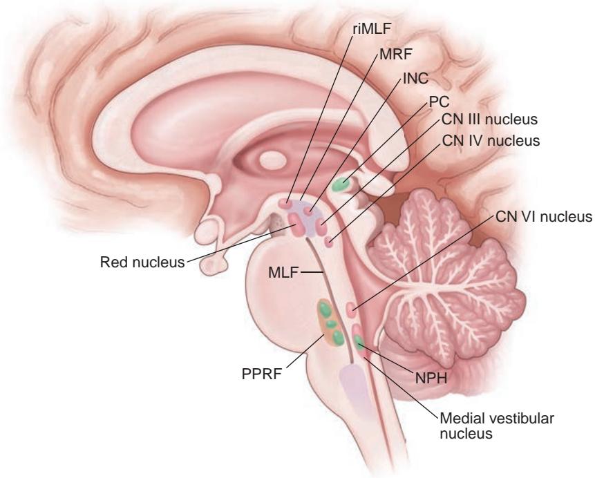
Figure 1-31 Schematic representation of a sagittal section of the brainstem showing the loca tion of the important structures involved in eye movements. The shaded area indicates the paramedian pontine reticular formation (PPRF). The dorsolateral pontine nucleus (DLPN) is not visible because this illustration is a midsagittal section, and the DLPN is laterally located; it is best visualized on an axial view through the rostral pons. CN = cranial nerve; INC = interstitial nucleus of Cajal; MLF = medial longitudinal fasciculus; MRF = mesencephalic reticular forma tion; NPH = nucleus prepositus hypoglossi; PC = posterior commissure; riMLF = rostral intersti tial nucleus of the MLF. (Illustration by Rob Flewell, CMI.)
dorsolateral pontine nuclei: neurons for smooth pursuit
nucleus prepositus hypoglossi-medial vestibular nucleus: neural integrator for hor izontal gaze
PPRF: excitatory burst neurons that generate horizontal saccades and inhibitory burst neurons that suppress horizontal saccades
CNs III, IV, and VI: neurons that project directly to EOMs
vestibular nuclei (CN VIII): neurons that project vestibular information to saccade generators and ocular motor CNs (Figs 1-32, 1-33)