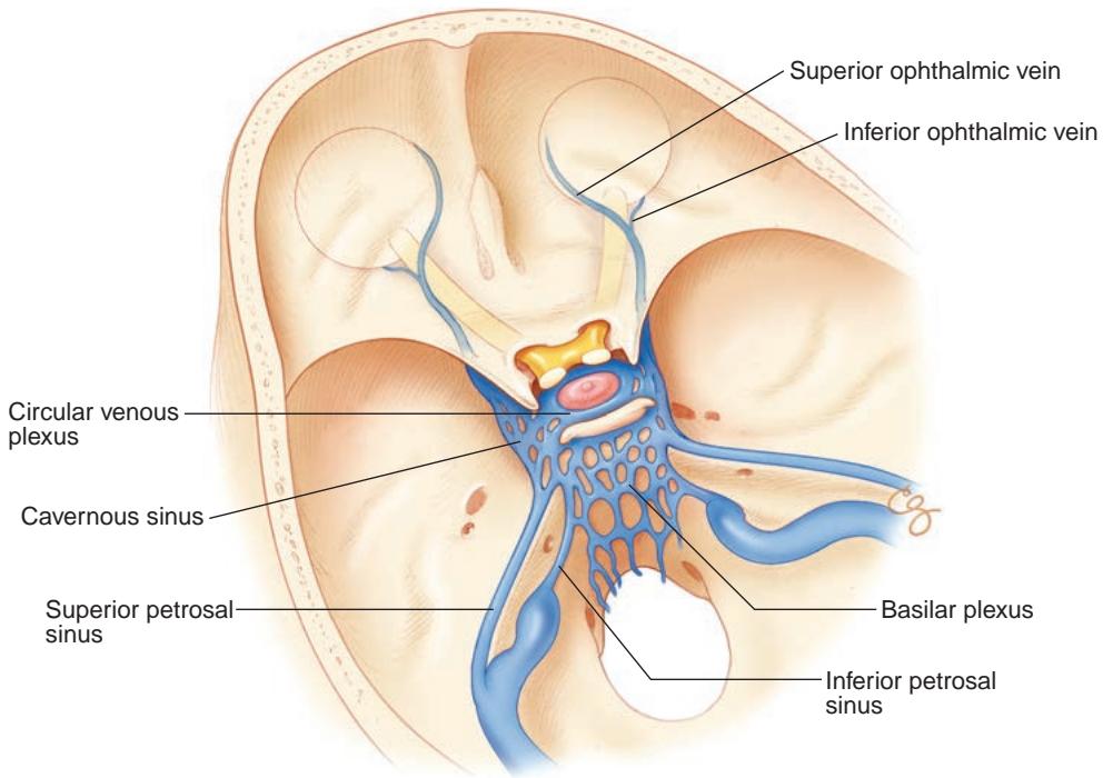
Figure 1-20 Anatomy of the cavernous sinus drainage system. (Illustration by Christine Gralapp.)
Veins of the eyelids anastomose medially between the angular vein and branches of the SOV in the superior medial orbit in the region of the trochlea. Facial veins drain in feriorly and laterally to form the external jugular vein, which eventually joins the internal jugular in the neck.
Lasjaunias P, Berenstein A, ter Brugge KG. Clinical Vascular Anatomy and Variations. 2nd ed. Springer; 2001. Surgical Neuroangiography; vol 1.
Afferent Visual Pathways
A disturbance in visual function may be the result of pathology affecting any part of the afferent visual system, including the retina, optic nerve, and visual pathways (Fig 1-21).
Retina
The afferent visual pathway begins within the retina. Details of retinal anatomy can be found in BCSC Section 2, Fundamentals and Principles of Ophthalmology, and Sec tion 12, Retina and Vitreous. The following discussion focuses on key points relevant to neuro-ophthalmology.
The absence of retinal receptors over the ONH creates a physiologic scotoma (blind spot), located approximately 15° from the fovea and measuring approximately 5° by 7°. The fovea, which is approximately 1.5 mm in diameter (1 disc diameter), is located about 4 mm (approximately 2.5 disc diameters) from and 0.8 mm lower than the ONH.
By contrast, the retinal pigment epithelium is in direct contact with the retinal pho toreceptor cells. Between the outer and inner retinal layers, the retinal signal generated in rods and cones is processed primarily through the bipolar cells that synapse on retinal ganglion cells (RGCs). A subset of melanopsin-containing RGCs, known as intrinsically photosensitive retinal ganglion cells (ipRGCs), serve primarily nonvisual light-dependent functions such as the pupillary light reflex. Horizontal, amacrine, and interplexiform cells (which communicate horizontally between neighboring cells) permit signal processing within the retinal layers. The glial support cells—Müller cells and astrocytes—play an impor tant metabolic role.

Figure 1-21 Basal view of the brain showing the anterior and posterior visual pathways. (Illustra tion by Dave Peace.)
The ratio of photoreceptor cells to RGCs varies depending on the region of the retina. The ratio is highest in the periphery (at more than 1000:1) and lowest at the fovea (where an RGC may receive a signal from a single cone). The foveal bipolar cells and RGCs are displaced radially from the fovea, with the bipolar cells receiving input via long cone axons that make up the Henle layer. The radial nature of the Henle layer is responsible for the accumulation of fluid in the macula in a star-shaped pattern.
Optic Nerve
The optic nerve begins anatomically at the ONH but physiologically and functionally within the RGC layer that covers the entire retina. Although ganglion cell fibers coming from the nasal retina can travel uninterrupted directly to the ONH, those coming from the temporal retina must anatomically separate to enter the ONH at either the superior or the inferior pole in order to avoid the macula (Fig 1-22). This unique anatomy means that some of the nasal fibers (nasal within the macula) enter the ONH on its temporal side (within the papillomacular bundle). Defects in the nerve fiber layer include focal loss, which may appear as either grooves or slits or as reflections that parallel the retinal arte rioles where the internal limiting membrane drapes over the vessels. Diffuse loss of the nerve fiber layer is often more difficult to detect and brings the retinal vessels into sharp relief.
The first portion of the optic nerve, representing the confluence of approximately 1.0–1.2 million RGC axons, traverses the sclera through the lamina cribrosa, which con tains approximately 200–300 channels. The combination of small channels and a unique blood supply (largely from branches of the posterior ciliary arteries) likely plays a role in several optic neuropathies. Anterograde and retrograde axonal transport of molecules, subcellular organelles, and metabolic products occurs along the length of the optic nerve. The anterograde axonal transport system is further subdivided into slow, intermediate, and fast speeds. This energy-dependent system, which requires high concentrations of oxygen, is sensitive to ischemic, inflammatory, and compressive processes.
Just posterior to the sclera, the optic nerve acquires a dural sheath that is contigu ous with the periorbita of the optic canal and an arachnoid membrane that supports and protects the axons and is contiguous with the arachnoid of the subdural intracranial space through the optic canal. This arrangement permits circulation of CSF around the optic nerve up to the ONH. Just posterior to the lamina cribrosa, the optic nerve also acquires a myelin coating, which increases its diameter from 1.5 mm (ie, in the ONH) to approxi mately 3 mm (6 mm including the optic nerve sheath). The myelin investment is part of the membrane of oligodendrocytes that join the nerve posterior to the sclera.
The intraorbital optic nerve extends approximately 25–30 mm to the optic canal, a length that allows unimpeded globe rotation as well as axial shifts within the orbit. The CRA and CRV travel within the anterior 10–12 mm of the optic nerve. The CRA supplies only a minor portion of the optic nerve circulation; most of the blood supply comes from pial branches of the surrounding meninges, which are in turn supplied by small branches of the OphA (see Fig 1-12). An approximate topographic representation is maintained in the optic nerve. Peripheral retinal representation is found more peripherally, and the pap illomacular bundle travels temporally and increasingly centrally within the nerve.
B
A
D