
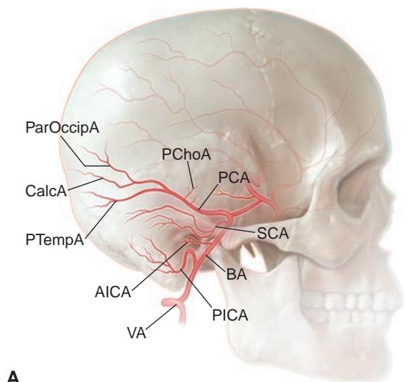
Figure 1-16 Vertebrobasilar arterial system and major arteries with common variations of the cortical branches of the posterior cerebral artery (PCA). A, Lateral view. B, Anteroposterior view. Vertebral artery (VA) and basilar artery (BA) branches: anterior inferior cerebellar artery (AICA); posterior inferior cerebellar artery (PICA); superior cerebellar artery (SCA). PCA and its branches: calcarine artery (CalcA); parieto-occipital artery (ParOccipA); posterior choroidal artery (PChoA); posterior temporal artery (PTempA); posterior communicating artery (PCoA). (Illustration by Rob Flewell, CMI.)
Venous System
Ocular venous outflow begins in the arcade retinal veins, which exit into the central retinal vein (CRV), and in the choroidal veins, which exit the sclera through the vortex veins. Ante riorly, the episcleral venous plexus collects both blood from the anterior uveal circulation and aqueous percolating through the Schlemm canal. These 3 primary venous drainage pathways (Fig 1-18) empty mainly into the superior ophthalmic vein (SOV), which runs posteriorly within the superior medial orbit to the orbital apex, where it crosses laterally to enter the cavernous sinus posterior to the superior orbital fissure. Cavernous sinus thrombosis or carotid-cavernous fistulas may cause venous congestion in the orbit and arterialization of the episcleral and conjunctival vessels (see Chapter 8, Fig 8-13).
Microscopic collaterals exist at variable levels between these venous beds. In rare in stances, shunts connecting retinal veins to choroidal veins are present within the retina. More commonly, optociliary shunt vessels (retinochoroidal collateral vessels) may appear on the ONH surface, usually with diseases that impair venous outflow, such as CRV oc clusion, optic nerve sheath meningioma, or optic nerve gliomas. At a more macroscopic level, the SOV is inconstantly connected anteriorly to the angular and facial veins and infe riorly to the inferior ophthalmic vein and pterygoid plexus. Venous collaterals are especially important in patients with elevated venous pressure due to a carotid-cavernous fistula and may provide endovascular access for embolization.

Figure 1-17 The occipital cortex and its blood supply. The primary visual cortex, or striate cortex (V1); the parastriate cortex (V2); and V3 are keyed by color. CalcA = calcarine artery; CC = corpus callosum; MCA = middle cerebral artery; PCA = posterior cerebral artery. (Illustration by Craig A. Luce.)
Intracranially, the superficial cortical venous system drains mainly superiorly and medially to the superior sagittal sinus running in the sagittal midline (Fig 1-19). In addi tion to facilitating cortical drainage, the superior sagittal sinus absorbs CSF through the arachnoid villi and the pacchionian granulations. The superior sagittal sinus continues posteriorly to terminate at the torcular Herophili (confluence of the venous sinuses) at the level of the tentorium that separates the cerebellum from the occipital lobes. Obstruction to venous outflow from venous sinus stenosis or thrombosis decreases CSF absorption and elevates intracranial pressure and may present as papilledema and/or CN VI palsy. From the point where the tentorium connects to the skull, the transverse sinuses run ante riorly to the petrous pyramid. Here, they turn to run caudally as the sigmoid sinus down to the jugular bulb, where the internal jugular vein exits the skull.
Inferior superficial cortical venous drainage is carried directly down to the trans verse and sigmoid sinuses through the vein of Labbé and the basilar vein of Rosenthal. Supratentorial diencephalon and mesencephalon drainage begins with the deep-draining veins (often in relation to the ventricular system). These deep veins merge to form the vein of Galen, which drains posteriorly into the straight sinus. Along with the superior sagittal sinus, the vein of Galen then runs within the tentorium to drain into the torcular Herophili.
Some anterior cerebral venous drainage may access the 2 cavernous sinuses, which are joined by variable connections through the sella and posteriorly through a plexus of veins over the clivus. The cavernous sinus drains primarily caudally into the jugular bulb via the inferior petrosal sinus, which traverses the Dorello canal with CN VI under the petroclinoid ligament. Alternatively, drainage may occur laterally along the petrous apex through the superior petrosal sinus to the junction of the transverse and sigmoid sinuses (Fig 1-20). Small veins may drain through the foramen rotundum and foramen ovale as well as through the pterygoid plexus to anastomose with the facial venous system (external jugular vein).
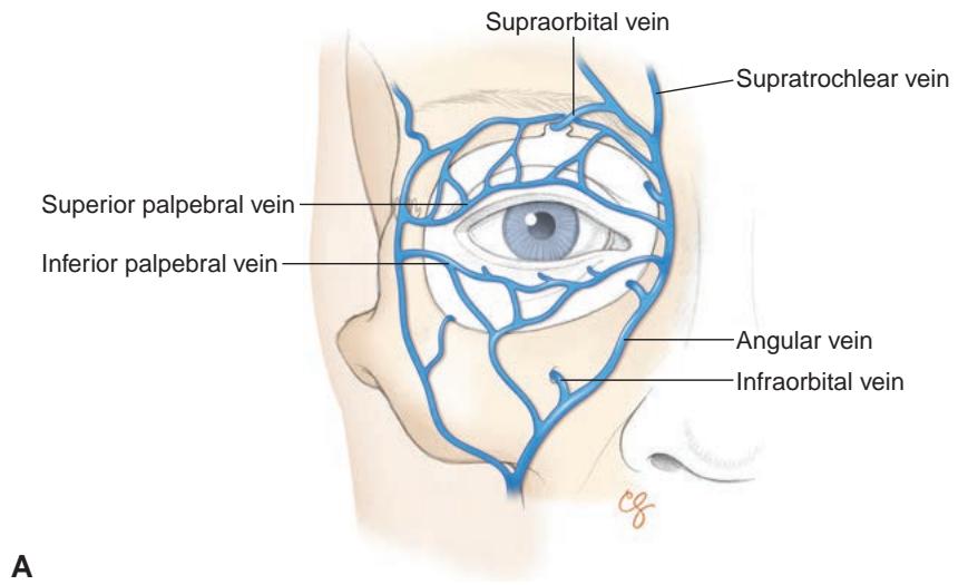

Figure 1-18 Venous drainage of the orbit. A, Anterior view of the superficial venous system of the eyelids. B, Sagittal view of the venous circulation of the orbit and globe. (Illustrations by Christine Gralapp.)
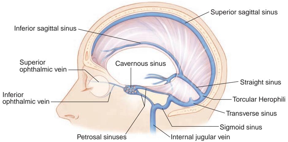
Figure 1-19 Anatomy of the cerebral venous sinus system. (Illustration by Christine Gralapp.)