When distal ACA occlusion occurs, the afferent visual pathways are spared; however, the premotor areas of the frontal lobes responsible for initiating saccades are supplied by branches of the ACA. Thus, patients with acute occlusion of the distal ACA may have a transient gaze preference and difficulty initiating contralateral saccades, although this ef fect is more commonly seen in patients with MCA territory lesions.

Figure 1-13 Relationship of the lateral geniculate nucleus (LGN) to nearby structures and its blood supply. AChoA = anterior choroidal artery; BC = brachium conjunctivum; CerePed = cere bral peduncle; CN = cranial nerve; ICA = internal carotid artery; MCA = middle cerebral artery; MGN = medial geniculate nucleus; ON = optic nerve; PCA = posterior cerebral artery; PCoA = pos terior communicating artery; PLChA = posterior lateral choroidal artery; Pulv = pulvinar; RN = red nucleus; SC = superior colliculus; SCA = superior cerebellar artery. (Illustration by Craig A. Luce.)
The MCA divides into several branches that supply the temporal lobe, parietal lobe, and superficial portions of the frontal and occipital lobes. Branches important to the vi sual pathways include those supplying the optic radiations as they traverse the deep white matter of the parietal and temporal lobes. Terminal branches of the MCA also variably supply the occipital tip representing the macula. This supply is chiefly responsible for the perimetric finding of macular sparing in PCA or calcarine artery occlusion (see Chap ter 5). In addition to supplying the afferent pathways, the MCA supplies the middle tem poral region, which is involved in visually guided pursuit movements.
CLINICAL PEARL
An MCA infarct causes ipsilateral pursuit dysfunction or asymmetry in optoki netic nystagmus (OKN) when a clinician rotates the OKN drum toward the side of the infarction.
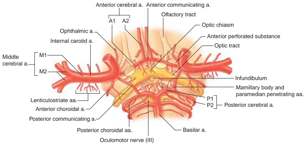
Figure 1-14 The circle of Willis represents an anastomosis of the anterior, middle, and poste rior cerebral arteries. Branches from these vessels supply the distal segment of the intracranial optic nerves, optic chiasm, and optic tract. a. = artery; aa = arteries. (Modified with permission from Liu GT, Volpe NJ, Galetta SL. Neuro-Ophthalmology: Diagnosis and Management. 2nd ed. Elsevier; 2010:295.)
Posterior Circulation
The posterior neurovascular circulation begins with the aortic arch. Beyond the origin of the common carotid artery, the innominate artery becomes the right subclavian artery. The left subclavian artery comes directly off the aorta. The right and left VAs subsequently arise from the subclavian arteries (see Fig 1-8A). The VA travels through a series of fo ramina in the lateral aspects of the cervical vertebral processes. After penetrating the dura at the foramen magnum, the VA gives rise to the posterior inferior cerebellar artery (PICA) before joining the other VA to form the BA (Fig 1-16). The PICA represents the more caudal of the 2 major circumferential arteries that wrap around the brainstem. Proximally, the PICA and BA give off branches that perforate the medial portion of the brainstem at the medullary level, followed by paramedian branches that supply the lateral aspects of the brainstem. Distally, the PICA supplies the inferior cerebellum, which is intimately involved in eye movements. VA or PICA occlusion is associated with lateral medullary syndrome (also called Wallenberg syndrome), which may present with ocular findings of Horner syndrome and ocular tilt reaction (see Chapter 9).
The second major circumferential arteries are branches of the anterior inferior cerebellar artery (AICA). The AICA arises from the more rostral BA and supplies the pontomedullary junction and distal cerebellum. A large proximal branch of the AICA, the internal auditory artery, supplies the CN VIII complex in the subarachnoid space and extends into the internal auditory canal. Along the course of the BA, small perforators arise directly to supply portions of the pons and midbrain. Median perforators of the BA supply the following structures:
the MLF
the paramedian pontine reticular formation (PPRF)
the medially located nuclei of CNs III, IV, and VI

Figure 1-15 Vascular supply of the optic nerve and visual pathway. (Modified with permission from Forrester JV, Dick AD, McMenamin PG, Roberts F, Pearlman E. The Eye: Basic Sciences in Practice. 4th ed. Elsevier; 2016:98.)
Interruption of these branches (which occurs commonly with vertebrobasilar atheroscle rotic disease or emboli) produces variable ophthalmoplegia, internuclear ophthalmople gia, and skew deviation. Pontine branches of the BA also supply the proximal portions of the CNs (particularly CN V) as they exit the brainstem.
The distal 2 sets of circumferential arteries consist of the superior cerebellar artery (SCA) followed by the PCA, representing the terminal branches of the BA at the level of the midbrain. Perforators from the proximal SCA partially supply the nucleus of CN III and its fascicles. In addition, small branches often supply the CN V root. CN III exits be tween the SCA and the PCA, where it may be compressed by an aneurysm.
Perforators from the proximal PCA (the P1 segment) supply the rostral portion of the midbrain (involved in vertical gaze) and part of the LGN. A large branch, the artery of Percheron, often supplies both sides of the midbrain from one of the PCAs. Because thalamostriate arteries originate from the P1 segment, infarcts related to the ICA–MCA spare the thalamus. The P1 segment ends with the PCoA, which joins the vertebrobasilar circulation to the carotid circulation anteriorly. The connecting PCoA parallels the course of CN III and then joins the ICA, which explains the occurrence of CN III palsy with PCoA aneurysms. As the distal PCA courses around the brainstem, it gives off a parieto- occipital branch before terminating in the calcarine branch, which supplies the primary visual cortex (Fig 1-17).