Сосудистая анатомия
Vascular Anatomy
Артериальная система
Arterial System
Ишемия является распространенной патофизиологической причиной зрительной дисфункции, включая двоение в глазах и потерю зрения. Знание сосудистой анатомии глаза имеет решающее значение для понимания возможности возникновения и купирования повреждений, вторичных по отношению к ишемии. Общие сонные артерии, отходящие от плечеголовного ствола справа и непосредственно от аорты слева, обеспечивают большую часть кровоснабжения черепа и его содержимого. Остальная часть кровоснабжения черепа осуществляется двумя позвоночными артериями (VAs), которые входят через большое затылочное отверстие после прохождения через отверстия в сегментах шейных позвонков. После проникновения VAs в твердую мозговую оболочку они соединяются в области мостомозгового перехода, формируя базилярную артерию (BA), которая поднимается вдоль передней поверхности моста и заканчивается двумя задними мозговыми артериями (PCAs) на уровне среднего мозга (рис. 1-8).
Ischemia is a common pathophysiologic cause of visual dysfunction, including double vision and loss of vision. Knowledge of the vascular anatomy of the eye is crucial in un derstanding the potential for and managing damage secondary to ischemia. The common carotid arteries, arising from the innominate artery on the right and directly from the aorta on the left, supply most of the blood to the skull and its contents. The remainder of the blood supply to the skull comes from the 2 vertebral arteries (VAs), which enter through the foramen magnum after traversing foramina in the cervical vertebral segments. Once the VAs penetrate the dura, they join near the pontomedullary junction to form the basilar artery (BA), which ascends along the anterior surface of the pons and terminates in the 2 posterior cerebral arteries (PCAs) at the level of the midbrain (Fig 1-8).
A
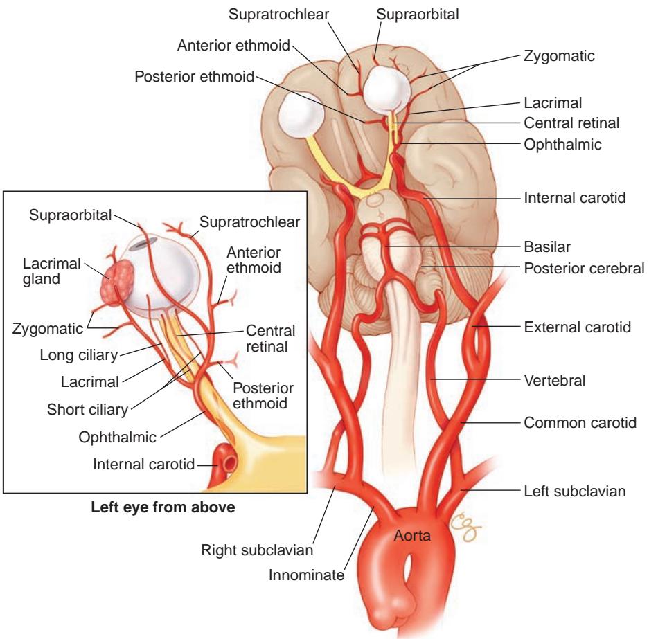
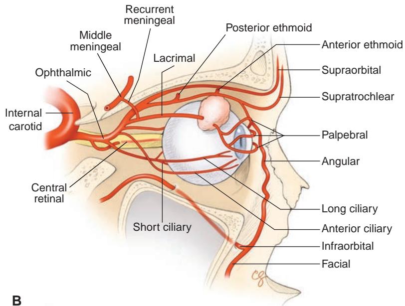
Рисунок 1-8 Артериальная карта орбиты и придатков глаза. A, Артериальное кровоснабжение орбиты и придатков глаза, берущее начало от дуги аорты. B, Сагиттальный вид артериального кровоснабжения орбиты и глазного яблока. (Иллюстрации выполнены Christine Gralapp.)
Figure 1-8 Arterial map of the orbit and ocular adnexa. A, Arterial supply of the orbit and ocular adnexa originating from the aortic arch. B, Sagittal view of the arterial supply to the orbit and globe. (Illustrations by Christine Gralapp.)
Передний отдел кровообращения
Anterior Circulation
Общая сонная артерия делится на наружную и внутреннюю ветви в области угла нижней челюсти (рис. 1-9). Наружная сонная артерия (ECA) обеспечивает кровоснабжение лица через свои основные ветви — лицевые артерии. Кожа головы снабжается кровью через ветви поверхностной височной артерии спереди и затылочной артерии сзади. Параназальные пазухи получают кровоснабжение из ветвей верхнечелюстной артерии (клиновидно-нёбной и подглазничной), которая заканчивается в крылонёбной ямке. Оболочки головного мозга снабжаются ветвями средней менингеальной артерии — основной ветви верхнечелюстной артерии, которая входит в среднюю черепную ямку через foramen spinosum, латеральнее foramen ovale. Ветви средней менингеальной артерии кровоснабжают параселлярную область, включая латеральную стенку кавернозного синуса, и заканчиваются в артериях круглого и овального отверстий. Различные менингеальные ветви могут входить в верхнюю глазничную щель. Терминальные ветви лицевой артерии снабжают кровью краевые аркады век. Для более подробного обсуждения кровоснабжения век см. BCSC Раздел 7, Oculofacial Plastic and Orbital Surgery.
The common carotid artery divides into external and internal branches at the angle of the jaw (Fig 1-9). The external carotid artery (ECA) supplies blood to the face through its major branches of the facial artery. The scalp is supplied via branches of the superficial tempo ral artery anteriorly and the occipital artery posteriorly. The paranasal sinuses receive their blood supply from branches of the maxillary artery (sphenopalatine and infraorbital), which terminates in the pterygopalatine fossa. The coverings of the brain are supplied by branches of the middle meningeal artery—a major branch of the maxillary artery—which enters the middle cranial fossa through the foramen spinosum, lateral to the foramen ovale. Branches of the middle meningeal artery supply the parasellar area, including the lateral wall of the cavernous sinus, and terminate in the arteries of the foramen rotundum and ovale. Variable meningeal branches may enter the superior orbital fissure. Terminal branches of the facial artery supply the marginal arcades of the eyelids. For further discussion of the blood supply of the eyelids, see BCSC Section 7, Oculofacial Plastic and Orbital Surgery.
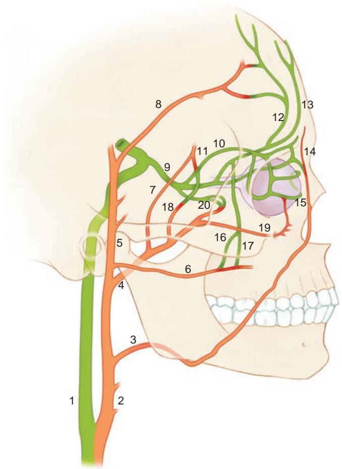
Рисунок 1-9 Коллатеральная анатомия внутренней сонной артерии (зеленый) и наружной сонной артерии (оранжевый). 1, внутренняя сонная; 2, наружная сонная; 3, лицевая; 4, верхнечелюстная; 5, поверхностная височная; 6, поперечно-лицевая; 7, средняя менингеальная; 8, лобная ветвь поверхностной височной; 9, глазничная; 10, слезная; 11, возвратная менингеальная; 12, надбровная; 13, надблоковая; 14, угловая; 15, вековая; 16, скуловисочная; 17, скулолицевая; 18, глубокая височная; 19, подглазничная; 20, мышечная. (Модифицировано с разрешения Kline LB. Neuro-Ophthalmology Review Manual. 6th ed. Slack; 2008:213. Иллюстрация Christine Gralapp.)
Figure 1-9 Internal carotid artery (green) and external carotid artery (orange) collateral anatomy. 1, internal carotid; 2, external carotid; 3, facial; 4, maxillary; 5, superficial temporal; 6, transverse facial; 7, middle meningeal; 8, frontal branch of superficial temporal; 9, oph thalmic; 10, lacrimal; 11, recurrent meningeal; 12, supraorbital; 13, supratrochlear; 14, angular; 15, palpebral; 16, zygomaticotemporal; 17, zygomaticofacial; 18, deep temporal; 19, infraor bital; 20, muscular. (Modified with permission from Kline LB. Neuro-Ophthalmology Review Manual. 6th ed. Slack; 2008:213. Illustration by Christine Gralapp.)
Крайне важно понимать степень коллатеральных связей между ветвями ECA и ICA (см. рис. 1-9). Интервенционные нейрорадиологи и нейрохирурги могут непреднамеренно эмболизировать дистальные ветви ICA, включая центральную артерию сетчатки (CRA), при введении частиц в ECA. Эмболизация наиболее вероятна при лечении артериовенозных мальформаций, но она также может произойти при эмболизации опухолей основания черепа перед хирургической резекцией.
It is extremely important to understand the extent of the collateral connections be tween the branches of the ECA and the ICA (see Fig 1-9). Interventional neuroradiolo gists and neurosurgeons may inadvertently embolize distal ICA branches, including the central retinal artery (CRA), while placing particles into the ECA. Embolization is most likely to occur during the treatment of arteriovenous malformations, but it can also occur when skull-base tumors are embolized before surgical resection.
Наиболее важные коллатеральные связи между наружным и внутренним кругами кровообращения проходят через орбиту, включая следующие артерии:
The most important collateral connections between the external and internal circula tions traverse the orbit, including the following arteries:
передняя и задняя решетчатые артерии медиально
anterior and posterior ethmoidal arteries medially
лицевая артерия в месте ее соединения с дистальными ветвями надблоковой и подблоковой артерий вокруг угловой артерии медиально
facial artery where it joins the distal branches of the supratrochlear and infratroch lear arteries around the angular artery medially
подглазничная и надбровная артерии (включая дистальные связи со слезной артерией) спереди
infraorbital and supraorbital arteries (including distal connections to the lacrimal artery) anteriorly
скуловисочная ветвь латерально
zygomaticotemporal branch laterally
различные твердооболочечные ветви, проходящие через верхнюю и нижнюю глазничные щели
various dural branches that traverse the superior and inferior orbital fissures
В редких случаях OphA может также отходить как ветвь менингеальной системы ECA.
In rare instances, the OphA may also arise as a branch of the meningeal system of the ECA.
ICA обеспечивает основной приток крови к внутричерепным структурам (рис. 1-10). ICA входит в череп через сонный канал. В пределах пирамиды височной кости артерия проходит рядом со средним и внутренним ухом, а также интрапетрозальным отделом CN VII. По достижении параселлярной области ICA поворачивает вверх непосредственно над рваным отверстием. Затем она входит в кавернозный синус, где сначала отдает менингогипофизарный ствол, а затем поворачивает вперед, проходя горизонтально параллельно телу клиновидной кости, чтобы отдать нижнелатеральный ствол, который образует анастомозы с ветвями средней менингеальной артерии. Ветви менингогипофизарного ствола кровоснабжают твердую оболочку задней части кавернозного синуса, а также CN III, IV, V и VI, входящие в кавернозный синус. Эти ветви также вариабельно кровоснабжают латеральный аспект турецкого седла, включая капсулу гипофиза и значительную часть самого гипофиза.
The ICA carries the major blood supply to the intracranial contents (Fig 1-10). The ICA enters the skull through the carotid canal. Within the petrous bone, the artery is near the middle and inner ear, as well as the intrapetrosal portion of CN VII. As the ICA reaches the parasellar area, it turns superiorly just above the foramen lacerum. It then enters the cavernous sinus, where it first gives off the meningohypophyseal trunk and then turns anteriorly to run horizontally parallel to the body of the sphenoid to give off the in ferolateral trunk, which forms anastomoses with branches of the middle meningeal artery. Branches of the meningohypophyseal trunk supply the dura of the posterior cavernous sinus as well as CNs III, IV, V, and VI entering the cavernous sinus. These branches also variably supply the lateral aspect of the sella turcica, including the pituitary capsule and a large portion of the pituitary gland itself.
В переднем отделе кавернозного синуса ICA делает петлю, меняя направление под передним клиновидным отростком и зрительным нервом. Эта петля проходит через два твердооболочечных кольца, оба расположенных близко к переднему клиновидному отростку (терминальной части малой крыши клиновидной кости). Проходя через второе кольцо, ICA становится интрадуральной (рис. 1-11). Вскоре после этого от ICA отходит глазничная артерия (OphA), которая входит в орбиту вместе со зрительным нервом через зрительный канал. Определение того, является ли аневризма ICA интрадуральной или экстрадуральной, имеет решающее значение, так как первая несет в себе внутренний риск жизнеугрожающего субарахноидального кровоизлияния при разрыве, тогда как вторая связана с низкой заболеваемостью при разрыве.
At the anterior aspect of the cavernous sinus, the ICA loops to reverse its direction under the anterior clinoid and the optic nerve. This loop passes through 2 dural rings, both close to the anterior clinoid (the terminal portion of the lesser wing of the sphenoid). As the ICA passes through the second ring, it becomes intradural (Fig 1-11). Shortly thereafter, the ICA gives off the OphA, which enters the orbit along with the optic nerve through the optic canal. Determining whether an ICA aneurysm is intradural or extradu ral is critical, as the former carries an inherent risk of life-threatening subarachnoid hem orrhage with rupture, whereas the latter is associated with low morbidity with rupture.
В пределах орбиты OphA (см. рис. 1-9) может анастомозировать с возвратными менингеальными ветвями, входящими через верхнюю глазничную щель. От OphA отходит CRA, которая затем входит в вещество зрительного нерва примерно в 10–12 мм кзади от глазного яблока. Внутри глаза CRA делится на верхнюю и нижнюю аркады. Эти
Within the orbit, the OphA (see Fig 1-9) may anastomose with recurrent meningeal branches that enter through the superior orbital fissure. The OphA gives off the CRA, which then enters the substance of the optic nerve approximately 10–12 mm posterior to the globe. Within the eye, the CRA divides into superior and inferior arcades. These
A
B