Вестибуло-окулярная система (вестибуло-окулярный рефлекс)
Vestibular ocular system (vestibular ocular reflex)
Вестибулярные ядра обеспечивают основной супрануклеарный вход для управления глазодвигательной функцией и тонический вход для позиционирования глаз.
The vestibular nuclei provide major supranuclear input into ocular motility and tonic input into eye positioning.
Вестибулярный нерв и улиточный нерв образуют комплекс CN VIII и выходят из пирамидной части височной кости через внутренний слуховой проход.
The vestibular nerve and cochlear nerve form the CN VIII complex and exit the petrous bone through the internal auditory meatus.
CN VIII проходит через субарахноидальное пространство в области мосто-мозжечкового угла к медиальному, латеральному и верхнему вестибулярным ядрам в продолговатом мозге.
CN VIII traverses the subarachnoid space within the cerebellopontine angle to me dial, lateral, and superior vestibular nuclei in the medulla.
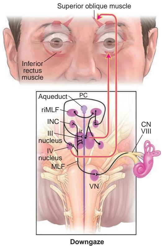

Рисунок 1-32 Анатомические схемы синтеза вертикальных движений глаз. От вертикальных полукружных каналов первичные афференты от CN VIII синаптируют с VN и поднимаются в MLF и соединительный тракт (brachium conjunctivum, не показан) к ядрам CN III и CN IV и INC. Возбуждающие пачечные нейроны в ростральном интерстициальном ядре MLF (riMLF) проецируются на мотонейроны CN III и IV и посылают коллатерали аксонов в INC. Ядро III = ядро глазодвигательного нерва (CN III); ядро IV = ядро блокового нерва (CN IV); PC = задняя спайка. (Иллюстрация Rob Flewell, CMI.)
Figure 1-32 Anatomical schemes for the synthesis of vertical eye movements. From the vertical semicircular canals, primary afferents from CN VIII synapse on the vestibular nuclei (VN) and ascend into the medial longitudinal fasciculus (MLF) and brachium conjunctivum (not shown) to the CN III and CN IV nuclei and the interstitial nucleus of Cajal (INC). Excitatory burst neurons in the rostral interstitial nucleus of the MLF (riMLF) project to the motoneurons of CNs III and IV and send axon collaterals to the INC. III nucleus = oculomotor nerve (CN III) nucleus; IV nucleus = trochlear nerve (CN IV) nucleus; PC = posterior commissure. (Illustration by Rob Flewell, CMI.)
Медиальное и нижнее вестибулярные ядра проецируются на нодулюс (центральное ядро мозжечка) и вентральную часть увулы.
The medial and inferior vestibular nuclei project to the nodulus (a central nucleus of the cerebellum) and the ventral uvula.
Информацию о клинических расстройствах вестибуло-окулярной функции см. в главе 9.
For information on clinical disorders of vestibular-ocular function, see Chapter 9.
Глазодвигательные черепные нервы
Ocular Motor Cranial Nerves
При отсутствии нейронной активности зрительные оси обычно умеренно или слабо дивергируют. Основной тонический вход для глазодвигательной функции обеспечивается 3 парами глазодвигательных CN — III, IV и VI, которые иннервируют 6 EOM, контролирующих движение глаз (рис. 1-34). Кроме того, CN III иннервирует верхний подниматель верхнего века и мышцы сфинктера зрачка.
Without neural activity, the visual axes are usually mildly to moderately divergent. The major tonic input to ocular motility is supplied by 3 pairs of ocular motor CNs—III, IV, and VI—that innervate the 6 EOMs controlling ocular movement (Fig 1-34). In addition, CN III innervates the levator palpebrae superioris and the pupillary sphincter muscles.
За исключением нижней косой мышцы, иннервация EOMs происходит примерно на одной трети расстояния от верхушки. Нижняя косая мышца получает иннервацию примерно в своей средней точке от нейроваскулярного пучка, проходящего параллельно латеральному аспекту нижней прямой мышцы. За исключением верхней косой мышцы, все EOMs получают иннервацию на внутренней поверхности. Ветви CN IV заканчиваются на верхней (наружной) поверхности мышцы.
Except for the inferior oblique muscle, the innervation of the EOMs occurs at ap proximately one-third of the distance from the apex. The inferior oblique muscle receives innervation at approximately its midpoint from a neurovascular bundle running parallel to the lateral aspect of the inferior rectus muscle. Except for the superior oblique muscle, all EOMs receive their innervation on the inside surface. The branches of CN IV termi nate on the upper (outer) surface of the muscle.

Рисунок 1-33 Анатомическая схема синтеза горизонтальных движений глаз. От горизонтального полукружного канала первичные афференты вестибулярного нерва (CN VIII) проецируются главным образом на нейроны ядра вестибулярного нерва (ядро VIII), которые затем посылают возбуждающий сигнал в контрлатеральное ядро CN VI. CN VI иннервирует ипсилатеральную наружную прямую мышцу и контрлатеральное ядро CN III через MLF. Горизонтальные саккады генерируются в фронтальном поле движений глаз, которое активирует контрлатеральную PPRF. Пачечные нейроны в PPRF стимулируют ипсилатеральное ядро CN VI, после чего путь идентичен пути, обеспечивающему горизонтальные движения глаз, генерируемые вестибулярным аппаратом. Некоторые нейроны в PPRF проецируются на ядро prepositus hypoglossi — медиальное вестибулярное ядро (нейронный интегратор), а затем в ядро CN VI. (Иллюстрация Rob Flewell, CMI.)
Figure 1-33 Anatomical scheme for the synthesis of horizontal eye movements. From the hori zontal semicircular canal, primary afferents of the vestibular nerve (CN VIII) project mainly to neurons of the vestibular nerve nucleus (VIII nucleus), which then send an excitatory connection to the contralateral CN VI nucleus. CN VI innervates the ipsilateral lateral rectus muscle and the contralateral CN III nucleus via the medial longitudinal fasciculus (MLF). Horizontal saccades are generated in the frontal eye field, which activates the contralateral paramedian pontine reticu lar formation (PPRF). Burst neurons in the PPRF stimulate the ipsilateral CN VI nucleus, with the subsequent pathway identical to that subserving horizontal vestibular-generated eye move ments. Some neurons in the PPRF project to the nucleus prepositus hypoglossi-medial vestibu lar nucleus (neural integrator) and then to the CN VI nucleus. (Illustration by Rob Flewell, CMI.)
Информацию о клинических проявлениях расстройств, вызванных инфрануклеарными, фасцикулярными и периферическими поражениями глазодвигательных CN, см. в главе 8.
See Chapter 8 for information on the clinical presentation of disorders due to infra- nuclear, fascicular, and peripheral ocular motor CN lesions.
Третий черепный нерв
Third cranial nerve
Ядро CN III (глазодвигательного нерва) расположено дорсально в среднем мозге, вентральнее водопровода, соединяющего третий и четвертый желудочки (рис. 1-35). Сам ядерный комплекс представляет собой совокупность подузлов, имеющих специфические идентифицируемые функции (рис. 1-36). Волокна, предназначенные для иннервации внутренней прямой, нижней прямой, нижней косой мышц, сфинктера зрачка и мышц цилиарного тела, выходят вентрально ипсилатерально от соответствующих ядер, из которых они происходят. Напротив, волокна из медиального подузла, расположенного вдоль срединной линии, перекрещиваются перед выходом из ствола мозга для иннервации контрлатеральной верхней прямой мышцы. Центральное хвостатое ядро обеспечивает двустороннюю иннервацию верхнего поднимателя верхнего века. Нуклеарное поражение CN III может вызвать ипсилатеральный паралич CN III с возможным дополнительным ограничением супрадукции контрлатерального глаза и/или двусторонним птозом в зависимости от того, какие подузлы поражены.
The CN III (oculomotor) nucleus is located dorsally within the midbrain ventral to the aqueduct connecting the third and fourth ventricles (Fig 1-35). The nuclear complex itself represents a collection of subnuclei that have specific identifiable functions (Fig 1-36). The fibers destined to innervate the medial rectus, inferior rectus, inferior oblique, pupil sphincter, and ciliary body muscles exit ventrally ipsilateral to the individual nuclei from which they originate. In contrast, the fibers from the medial subnucleus, which lies along the midline, cross before exiting the brainstem to innervate the contralateral superior rec tus muscle. The central caudate nucleus provides innervation to the levator palpebrae superioris bilaterally. A nuclear CN III lesion can cause an ipsilateral CN III palsy with possible additional limitation of supraduction of the contralateral eye and/or bilateral pto sis depending on which subnuclei are affected.
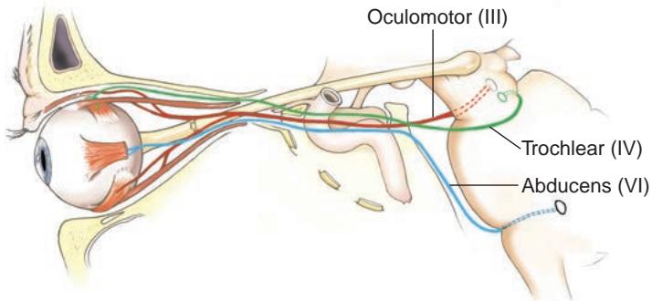
Рисунок 1-34 Вид сбоку на ход CN III, IV и VI. (Иллюстрация Dave Peace.)
Figure 1-34 Lateral view of the course of CNs III, IV, and VI. (Illustration by Dave Peace.)
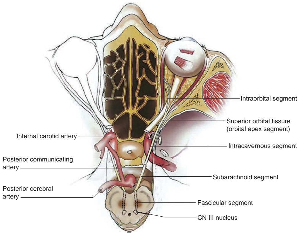
Рисунок 1-35 Иллюстрация аксиального хода CN III от ее начала в среднем мозге. (Перепечатано из Foroozan R, Bhatti MT, Rhoton AL. Transsphenoidal diplopia. Surv Ophthalmol. 2004;49(3):352; с разрешения Elsevier.)
Figure 1-35 Illustration of the axial course of CN III from its origin in the midbrain. (Reprinted from Foroozan R, Bhatti MT, Rhoton AL. Transsphenoidal diplopia. Surv Ophthalmol. 2004;49(3):352; with permission from Elsevier.)