
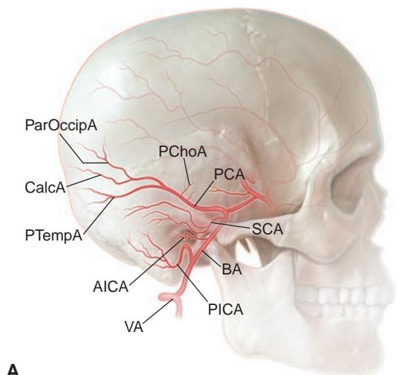
Рисунок 1-16 Вертебробазилярная артериальная система и основные артерии с распространенными вариантами корковых ветвей задней мозговой артерии (PCA). A, вид сбоку. B, переднезадний вид. Ветви позвоночной артерии (VA) и базилярной артерии (BA): anterior inferior cerebellar artery (AICA); posterior inferior cerebellar artery (PICA); superior cerebellar artery (SCA). PCA и ее ветви: валиковая артерия (CalcA); теменно-затылочная артерия (ParOccipA); задняя хориоидальная артерия (PChoA); задняя височная артерия (PTempA); задняя соединительная артерия (PCoA). (Иллюстрация Rob Flewell, CMI.)
Figure 1-16 Vertebrobasilar arterial system and major arteries with common variations of the cortical branches of the posterior cerebral artery (PCA). A, Lateral view. B, Anteroposterior view. Vertebral artery (VA) and basilar artery (BA) branches: anterior inferior cerebellar artery (AICA); posterior inferior cerebellar artery (PICA); superior cerebellar artery (SCA). PCA and its branches: calcarine artery (CalcA); parieto-occipital artery (ParOccipA); posterior choroidal artery (PChoA); posterior temporal artery (PTempA); posterior communicating artery (PCoA). (Illustration by Rob Flewell, CMI.)
Венозная система
Venous System
Венозный отток из глаза начинается в сетчаточных венах аркад, которые впадают в центральную вену сетчатки (CRV), и в хориоидальных венах, которые выходят из склеры через вортикозные вены. Спереди эписклеральное венозное сплетение собирает как кровь из переднего увеального круга, так и водянистую влагу, просачивающуюся через канал Шлемма. Эти 3 основных пути венозного дренажа (рис. 1-18) впадают главным образом в верхнюю глазничную вену (SOV), которая проходит сзади в верхне-медиальном отделе орбиты к верхушке орбиты, где она смещается латерально, чтобы войти в кавернозный синус позади верхней глазничной щели. Тромбоз кавернозного синуса или каротидно-кавернозные фистулы могут вызвать венозный застой в орбите и артериализацию эписклеральных и конъюнктивальных сосудов (см. главу 8, рис. 8-13).
Ocular venous outflow begins in the arcade retinal veins, which exit into the central retinal vein (CRV), and in the choroidal veins, which exit the sclera through the vortex veins. Ante riorly, the episcleral venous plexus collects both blood from the anterior uveal circulation and aqueous percolating through the Schlemm canal. These 3 primary venous drainage pathways (Fig 1-18) empty mainly into the superior ophthalmic vein (SOV), which runs posteriorly within the superior medial orbit to the orbital apex, where it crosses laterally to enter the cavernous sinus posterior to the superior orbital fissure. Cavernous sinus thrombosis or carotid-cavernous fistulas may cause venous congestion in the orbit and arterialization of the episcleral and conjunctival vessels (see Chapter 8, Fig 8-13).
Микроскопические коллатерали существуют на различных уровнях между этими венозными бассейнами. В редких случаях в сетчатке присутствуют шунты, соединяющие сетчаточные вены с хориоидальными венами. Чаще на поверхности ДЗН могут появляться оптоцилиарные шунты (ретинохориоидальные коллатеральные сосуды), обычно при заболеваниях, нарушающих венозный отток, таких как окклюзия CRV, менингиома оболочки зрительного нерва или глиомы зрительного нерва. На более макроскопическом уровне SOV непостоянно соединяется спереди с угловой и лицевой венами, а снизу с нижней глазничной веной и крыловидным сплетением. Венозные коллатерали особенно важны у пациентов с повышенным венозным давлением из-за каротидно-кавернозной фистулы и могут обеспечить эндоваскулярный доступ для эмболизации.
Microscopic collaterals exist at variable levels between these venous beds. In rare in stances, shunts connecting retinal veins to choroidal veins are present within the retina. More commonly, optociliary shunt vessels (retinochoroidal collateral vessels) may appear on the ONH surface, usually with diseases that impair venous outflow, such as CRV oc clusion, optic nerve sheath meningioma, or optic nerve gliomas. At a more macroscopic level, the SOV is inconstantly connected anteriorly to the angular and facial veins and infe riorly to the inferior ophthalmic vein and pterygoid plexus. Venous collaterals are especially important in patients with elevated venous pressure due to a carotid-cavernous fistula and may provide endovascular access for embolization.

Рисунок 1-17 Затылочная кора и ее кровоснабжение. Первичная зрительная кора, или полосатая кора (V1); парастриарная кора (V2); и V3 обозначены цветом. CalcA = валиковая артерия; CC = мозолистое тело; MCA = средняя мозговая артерия; PCA = задняя мозговая артерия. (Иллюстрация Craig A. Luce.)
Figure 1-17 The occipital cortex and its blood supply. The primary visual cortex, or striate cortex (V1); the parastriate cortex (V2); and V3 are keyed by color. CalcA = calcarine artery; CC = corpus callosum; MCA = middle cerebral artery; PCA = posterior cerebral artery. (Illustration by Craig A. Luce.)
Внутричерепно поверхностная корковая венозная система дренируется главным образом сверху и медиально в верхний сагиттальный синус, проходящий по сагиттальной срединной линии (рис. 1-19). Помимо обеспечения коркового дренажа, верхний сагиттальный синус абсорбирует CSF через арахноидальные ворсинки и грануляции Пакье. Верхний сагиттальный синус продолжается сзади и заканчивается в torcular Herophili (слиянии венозных синусов) на уровне намета мозжечка, который отделяет мозжечок от затылочных долей. Нарушение венозного оттока вследствие стеноза или тромбоза венозного синуса снижает абсорбцию CSF и повышает внутричерепное давление, что может проявляться отеком диска зрительного нерва и/или параличом CN VI. От места прикрепления намета мозжечка к черепу поперечные синусы идут вперед к пирамиде височной кости. Здесь они поворачивают и идут каудально как сигмовидный синус вниз к яремному булбусу, где внутренняя яремная вена выходит из черепа.
Intracranially, the superficial cortical venous system drains mainly superiorly and medially to the superior sagittal sinus running in the sagittal midline (Fig 1-19). In addi tion to facilitating cortical drainage, the superior sagittal sinus absorbs CSF through the arachnoid villi and the pacchionian granulations. The superior sagittal sinus continues posteriorly to terminate at the torcular Herophili (confluence of the venous sinuses) at the level of the tentorium that separates the cerebellum from the occipital lobes. Obstruction to venous outflow from venous sinus stenosis or thrombosis decreases CSF absorption and elevates intracranial pressure and may present as papilledema and/or CN VI palsy. From the point where the tentorium connects to the skull, the transverse sinuses run ante riorly to the petrous pyramid. Here, they turn to run caudally as the sigmoid sinus down to the jugular bulb, where the internal jugular vein exits the skull.
Нижний поверхностный корковый венозный дренаж осуществляется непосредственно в поперечные и сигмовидные синусы через вену Лаббе и базилярную вену Розенталя. Дренаж супратенториального диэнцефалона и мезенцефалона начинается с глубоких вен (часто в связи с вентрикулярной системой). Эти глубокие вены сливаются, образуя вену Галена, которая дренируется сзади в прямой синус. Вместе с верхним сагиттальным синусом вена Галена затем проходит внутри намета мозжечка, чтобы впасть в torcular Herophili.
Inferior superficial cortical venous drainage is carried directly down to the trans verse and sigmoid sinuses through the vein of Labbé and the basilar vein of Rosenthal. Supratentorial diencephalon and mesencephalon drainage begins with the deep-draining veins (often in relation to the ventricular system). These deep veins merge to form the vein of Galen, which drains posteriorly into the straight sinus. Along with the superior sagittal sinus, the vein of Galen then runs within the tentorium to drain into the torcular Herophili.
Часть переднего церебрального венозного дренажа может поступать в два кавернозных синуса, которые соединены вариабельными связями через турецкое седло и сзади через сплетение вен над скатом. Кавернозный синус дренируется преимущественно каудально в яремный булбус через нижний петрозальный синус, который проходит через канал Дорелло вместе с CN VI под петроклиноидной связкой. В качестве альтернативы дренаж может происходить латерально вдоль верхушки пирамиды височной кости через верхний петрозальный синус к месту слияния поперечного и сигмовидного синусов (рис. 1-20). Мелкие вены могут дренироваться через foramen rotundum и foramen ovale, а также через крыловидное сплетение, анастомозируя с лицевой венозной системой (наружная яремная вена).
Some anterior cerebral venous drainage may access the 2 cavernous sinuses, which are joined by variable connections through the sella and posteriorly through a plexus of veins over the clivus. The cavernous sinus drains primarily caudally into the jugular bulb via the inferior petrosal sinus, which traverses the Dorello canal with CN VI under the petroclinoid ligament. Alternatively, drainage may occur laterally along the petrous apex through the superior petrosal sinus to the junction of the transverse and sigmoid sinuses (Fig 1-20). Small veins may drain through the foramen rotundum and foramen ovale as well as through the pterygoid plexus to anastomose with the facial venous system (external jugular vein).
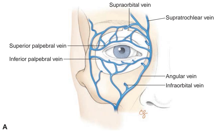

Рисунок 1-18 Венозный дренаж орбиты. A, вид спереди на поверхностную венозную систему век. B, сагиттальный вид венозного кровообращения орбиты и глазного яблока. (Иллюстрации Christine Gralapp.)
Figure 1-18 Venous drainage of the orbit. A, Anterior view of the superficial venous system of the eyelids. B, Sagittal view of the venous circulation of the orbit and globe. (Illustrations by Christine Gralapp.)
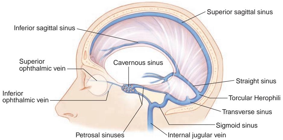
Рисунок 1-19 Анатомия системы венозных синусов головного мозга. (Иллюстрация Christine Gralapp.)
Figure 1-19 Anatomy of the cerebral venous sinus system. (Illustration by Christine Gralapp.)