Fifth Cranial Nerve
The sensory nerve, CN V (trigeminal nerve), terminates within the trigeminal nucleus. The nuclear complex of CN V extends from the midbrain to the cervical spinal cord.

Figure 1-40 Primary, secondary, and tertiary functions of the extraocular muscles, right eye. (Illustration by Christine Gralapp.)
| Muscle | Primary | Secondary | Tertiary |
| Medial rectus | Adduction | ||
| Lateral rectus | Abduction | ||
| Inferior rectus | Depression | Extorsion | Adduction |
| Superior rectus | Elevation | Intorsion | Adduction |
| Inferior oblique | Extorsion | Elevation | Abduction |
| Superior oblique | Intorsion | Depression | Abduction |
The following sensory nuclei are included within the CN V nuclear complex:
main sensory nucleus: located within the pons lateral to the motor nucleus; it re ceives light-touch information from the skin of the face and mucous membranes
mesencephalic nucleus: serves proprioception and deep sensation from the facial muscles, including the EOMs and the muscles of mastication (temporalis, ptery goid, and masseter)
spinal nucleus: extends caudally to the level of C4 vertebra; it receives pain and tem perature sensation and cutaneous sensory information
Damage to the spinal nucleus of CN V (eg, a brainstem lesion) causes bilateral sensory loss in concentric rings of the face, whereas peripheral sensory loss is unilateral and assumes the pattern of the ophthalmic, maxillary, or mandibular divisions of the trigeminal nerve (Fig 1-41).
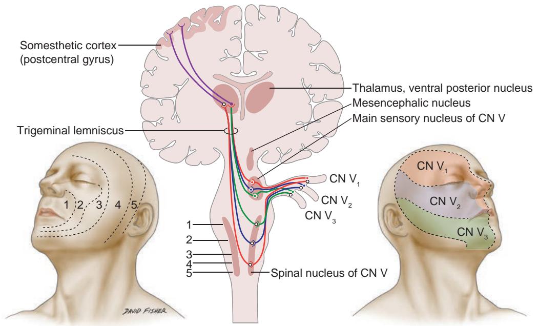
Figure 1-41 Diagram of the central pathways and peripheral innervation of CN V. The numbers 1–5 indicate the location of dermatomes on the face and their corresponding representation in the brainstem. (Used with permission from Kline LB. Neuro-Ophthalmology Review Manual. 6th ed. Slack; 2008:174. Illustration by David Fisher).
The various sensory nuclei of CN V project to the contralateral thalamus and from there to the postcentral gyrus. The oculocardiac reflex is mediated by the trigeminal nerve (afferent limb) and parasympathetic output of the vagus nerve (efferent limb).
Medial to the main sensory nucleus, the motor nucleus of CN V lies in the pons. The motor nucleus sends signals to the muscles of mastication, the tensor tympani (which re flexively dampens loud noises by tensing the tympanic membrane within the middle ear), the tensor veli palatini (which orients the uvula), the mylohyoid, and the anterior belly of the digastric muscle.
The fascicles of CN V enter the brainstem ventrally in the pons and extra-axially tra verse the subarachnoid space to penetrate the dura just over the petrous pyramid. Within the subarachnoid space, the trigeminal root often comes in contact with the SCA. This proximity may be a cause of trigeminal neuralgia (atypical facial pain, discussed in Chap ter 13) and is the anatomical basis for microvascular decompression. The 3 divisions of CN V synapse in the trigeminal (gasserian) ganglion (Fig 1-42), located in an extradural space at the floor of the middle cranial fossa (Meckel cave).
Ophthalmic division
The ophthalmic division (CN V1) is the most anterior branch of CN V exiting the trigeminal ganglion. It runs forward within the lateral wall of the cavernous sinus just below CN IV. As it approaches the superior orbital fissure extradurally, it divides into 3 major branches: lac rimal, frontal, and nasociliary. In addition, small branches innervate the dura of the anterior
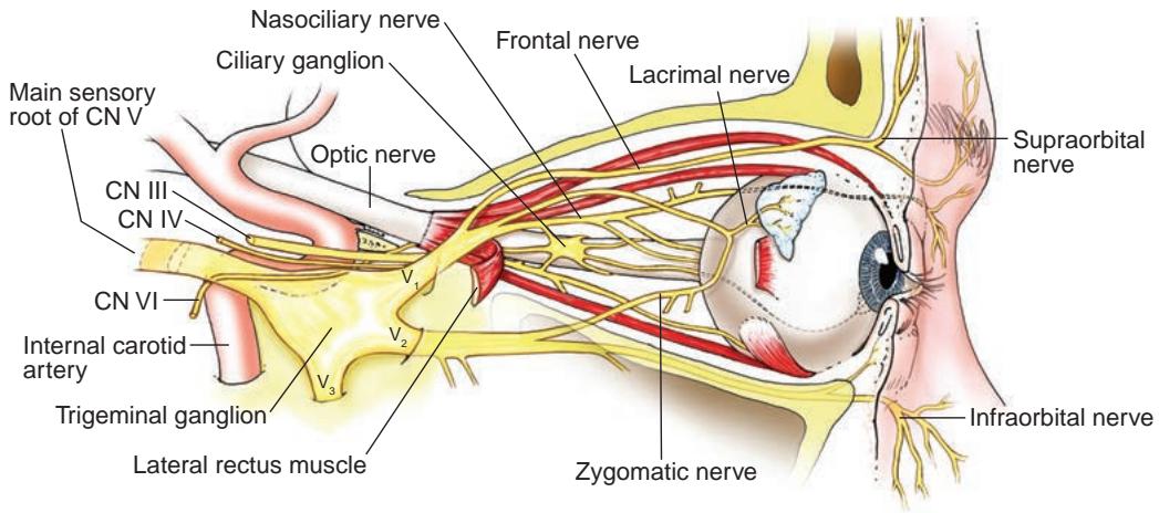
Figure 1-42 Lateral view of the orbit, showing its sensory nerves. (Illustration by Dave Peace.)
middle cranial fossa, including the cavernous sinus, the parasellar region, the tentorium, and the dura of the petrous apex. These branches also innervate the floor of the anterior cranial fossa, including the falx cerebri and the major blood vessels at the skull base.
The lacrimal and frontal nerves enter the orbital apex outside the annulus of Zinn. At its terminus, the frontal nerve divides into supraorbital and supratrochlear branches, which innervate the forehead, frontal sinus, and upper eyelid (including the conjunctiva). The lacrimal nerve also runs anteriorly in the superior lateral orbit slightly above the lat eral rectus to innervate the lacrimal gland and the skin just superotemporal to the orbit. The nasociliary nerve is the only branch that enters the intraconal space through the an nulus of Zinn. The nasociliary branch runs through the ciliary ganglion and anteriorly to innervate the globe through the short and long posterior ciliary nerves. Before reaching the globe, branches from the nasociliary division pass through the anterior and posterior ethmoidal foramina to innervate part of the ethmoid sinuses, the lateral wall of the nose, and the skin of the nose to the nasal tip. The co-innervation of the globe and the nasal skin explains the development of Hutchinson sign (vesicles on the tip or side of the nose) in patients with herpes zoster ophthalmicus.