Пятый черепной нерв
Fifth Cranial Nerve
Сенсорный нерв, CN V (тройничный нерв), заканчивается в тройничном ядре. Ядерный комплекс CN V простирается от среднего мозга до шейного отдела спинного мозга.
The sensory nerve, CN V (trigeminal nerve), terminates within the trigeminal nucleus. The nuclear complex of CN V extends from the midbrain to the cervical spinal cord.

Рисунок 1-40 Первичные, вторичные и третичные функции глазодвигательных мышц, правый глаз. (Иллюстрация Кристин Гралапп.)
Figure 1-40 Primary, secondary, and tertiary functions of the extraocular muscles, right eye. (Illustration by Christine Gralapp.)
| Мышца | Первичная | Вторичная | Третичная |
| Внутренняя прямая мышца | Аддукция | ||
| Наружная прямая мышца | Абдукция | ||
| Нижняя прямая мышца | Депрессия | Эксторсия | Аддукция |
| Верхняя прямая мышца | Элевация | Инторсия | Аддукция |
| Нижняя косая мышца | Эксторсия | Элевация | Абдукция |
| Верхняя косая мышца | Инторсия | Депрессия | Абдукция |
| Muscle | Primary | Secondary | Tertiary |
| Medial rectus | Adduction | ||
| Lateral rectus | Abduction | ||
| Inferior rectus | Depression | Extorsion | Adduction |
| Superior rectus | Elevation | Intorsion | Adduction |
| Inferior oblique | Extorsion | Elevation | Abduction |
| Superior oblique | Intorsion | Depression | Abduction |
В ядерный комплекс CN V входят следующие сенсорные ядра:
The following sensory nuclei are included within the CN V nuclear complex:
главное сенсорное ядро: расположено в мосту латеральнее двигательного ядра; оно получает информацию о легком прикосновении от кожи лица и слизистых оболочек
main sensory nucleus: located within the pons lateral to the motor nucleus; it re ceives light-touch information from the skin of the face and mucous membranes
мезенцефалическое ядро: обеспечивает проприоцепцию и глубокую чувствительность мышц лица, включая глазодвигательные мышцы и жевательные мышцы (височную, крыловидные и массетерную)
mesencephalic nucleus: serves proprioception and deep sensation from the facial muscles, including the EOMs and the muscles of mastication (temporalis, ptery goid, and masseter)
спинальное ядро: простирается каудально до уровня C4 позвонка; оно получает болевую и температурную чувствительность, а также кожную сенсорную информацию
spinal nucleus: extends caudally to the level of C4 vertebra; it receives pain and tem perature sensation and cutaneous sensory information
Повреждение спинального ядра CN V (например, при поражении ствола мозга) вызывает двустороннюю потерю чувствительности концентрическими кольцами на лице, тогда как периферическая потеря чувствительности является односторонней и соответствует распределению глазной, верхнечелюстной или нижнечелюстной ветвей тройничного нерва (рис. 1-41).
Damage to the spinal nucleus of CN V (eg, a brainstem lesion) causes bilateral sensory loss in concentric rings of the face, whereas peripheral sensory loss is unilateral and assumes the pattern of the ophthalmic, maxillary, or mandibular divisions of the trigeminal nerve (Fig 1-41).
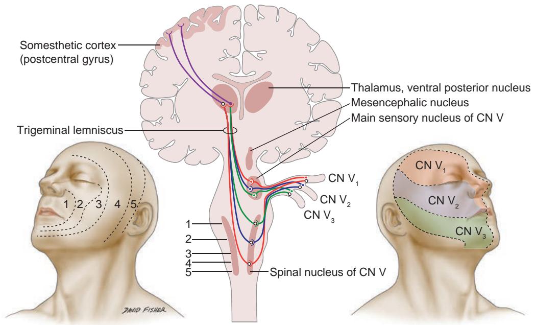
Рисунок 1-41 Схема центральных путей и периферической иннервации CN V. Цифры 1–5 указывают расположение дерматомов на лице и их соответствующее представительство в стволе мозга. (Использовано с разрешения Kline LB. Neuro-Ophthalmology Review Manual. 6th ed. Slack; 2008:174. Иллюстрация Дэвида Фишера).
Figure 1-41 Diagram of the central pathways and peripheral innervation of CN V. The numbers 1–5 indicate the location of dermatomes on the face and their corresponding representation in the brainstem. (Used with permission from Kline LB. Neuro-Ophthalmology Review Manual. 6th ed. Slack; 2008:174. Illustration by David Fisher).
Различные сенсорные ядра CN V проецируются на контралатеральный таламус и оттуда на постцентральную извилину. Окулокардиальный рефлекс опосредован тройничным нервом (афферентное звено) и парасимпатическим выходом блуждающего нерва (эфферентное звено).
The various sensory nuclei of CN V project to the contralateral thalamus and from there to the postcentral gyrus. The oculocardiac reflex is mediated by the trigeminal nerve (afferent limb) and parasympathetic output of the vagus nerve (efferent limb).
Медиальнее главного сенсорного ядра в мосту располагается двигательное ядро CN V. Двигательное ядро посылает сигналы к жевательным мышцам, напрягателю барабанной перепонки (который рефлекторно приглушает громкие звуки, напрягая барабанную перепонку в среднем ухе), напрягателю небной занавески (который ориентирует язычок), двубрюшной мышце (переднему брюшку) и подязычно-челюстной мышце.
Medial to the main sensory nucleus, the motor nucleus of CN V lies in the pons. The motor nucleus sends signals to the muscles of mastication, the tensor tympani (which re flexively dampens loud noises by tensing the tympanic membrane within the middle ear), the tensor veli palatini (which orients the uvula), the mylohyoid, and the anterior belly of the digastric muscle.
Пучки CN V входят в ствол мозга вентрально в области моста и экстрааксиально пересекают субарахноидальное пространство, чтобы проникнуть в твердую мозговую оболочку непосредственно над пирамидой височной кости. В субарахноидальном пространстве корень тройничного нерва часто контактирует с SCA. Эта близость может быть причиной тройничного невралгии (атипичная лицевая боль, обсуждается в главе 13) и является анатомической основой для микрососудистой декомпрессии. 3 ветви CN V синаптируют в тройничном (гассеровом) ганглии (рис. 1-42), расположенном в экстрадуральном пространстве на дне средней черепной ямки (пещера Меккеля).
The fascicles of CN V enter the brainstem ventrally in the pons and extra-axially tra verse the subarachnoid space to penetrate the dura just over the petrous pyramid. Within the subarachnoid space, the trigeminal root often comes in contact with the SCA. This proximity may be a cause of trigeminal neuralgia (atypical facial pain, discussed in Chap ter 13) and is the anatomical basis for microvascular decompression. The 3 divisions of CN V synapse in the trigeminal (gasserian) ganglion (Fig 1-42), located in an extradural space at the floor of the middle cranial fossa (Meckel cave).
Глазная ветвь
Ophthalmic division
Глазная ветвь (CN V1) является самой передней ветвью CN V, выходящей из тройничного ганглия. Она проходит вперед в латеральной стенке кавернозного синуса непосредственно под CN IV. При экстрадуральном приближении к верхней глазничной щели она делится на 3 основные ветви: слезную, лобную и носоресничную. Кроме того, мелкие ветви иннервируют твердую мозговую оболочку передней
The ophthalmic division (CN V1) is the most anterior branch of CN V exiting the trigeminal ganglion. It runs forward within the lateral wall of the cavernous sinus just below CN IV. As it approaches the superior orbital fissure extradurally, it divides into 3 major branches: lac rimal, frontal, and nasociliary. In addition, small branches innervate the dura of the anterior
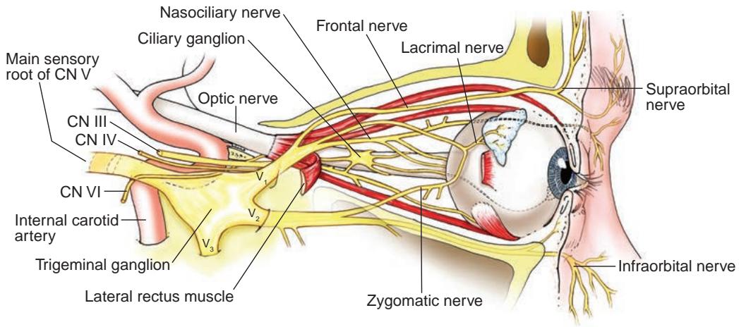
Рисунок 1-42 Вид орбиты сбоку, демонстрирующий ее сенсорные нервы. (Иллюстрация Дейва Писа.)
Figure 1-42 Lateral view of the orbit, showing its sensory nerves. (Illustration by Dave Peace.)
средней черепной ямки, включая кавернозный синус, параселлярную область, намета мозжечка и твердую мозговую оболочку верхушки пирамиды височной кости. Эти ветви также иннервируют дно передней черепной ямки, включая серп мозга и основные кровеносные сосуды у основания черепа.
middle cranial fossa, including the cavernous sinus, the parasellar region, the tentorium, and the dura of the petrous apex. These branches also innervate the floor of the anterior cranial fossa, including the falx cerebri and the major blood vessels at the skull base.
Слезный и лобный нервы входят в верхушку орбиты вне кольца Цинна. В своем окончании лобный нерв делится на надбровную и надблоковую ветви, которые иннервируют лоб, лобный синус и верхнее веко (включая конъюнктиву). Слезный нерв также проходит вперед в верхнелатеральной части орбиты чуть выше наружной прямой мышцы, чтобы иннервировать слезную железу и кожу непосредственно верхневисочно от орбиты. Носоресничный нерв является единственной ветвью, которая входит в интракональное пространство через кольцо Цинна. Носоресничная ветвь проходит через ресничный ганглий и вперед, чтобы иннервировать глазное яблоко через короткие и длинные задние ресничные нервы. Перед достижением глазного яблока ветви носоресничного отдела проходят через переднее и заднее решетчатые отверстия, чтобы иннервировать часть решетчатых синусов, латеральную стенку носа и кожу носа до кончика носа. Совместная иннервация глазного яблока и кожи носа объясняет развитие признака Хатчинсона (везикулы на кончике или боковой поверхности носа) у пациентов с опоясывающим лишаем глазной области (herpes zoster ophthalmicus).
The lacrimal and frontal nerves enter the orbital apex outside the annulus of Zinn. At its terminus, the frontal nerve divides into supraorbital and supratrochlear branches, which innervate the forehead, frontal sinus, and upper eyelid (including the conjunctiva). The lacrimal nerve also runs anteriorly in the superior lateral orbit slightly above the lat eral rectus to innervate the lacrimal gland and the skin just superotemporal to the orbit. The nasociliary nerve is the only branch that enters the intraconal space through the an nulus of Zinn. The nasociliary branch runs through the ciliary ganglion and anteriorly to innervate the globe through the short and long posterior ciliary nerves. Before reaching the globe, branches from the nasociliary division pass through the anterior and posterior ethmoidal foramina to innervate part of the ethmoid sinuses, the lateral wall of the nose, and the skin of the nose to the nasal tip. The co-innervation of the globe and the nasal skin explains the development of Hutchinson sign (vesicles on the tip or side of the nose) in patients with herpes zoster ophthalmicus.