CLINICAL PEARL
Lesions of the clivus can cause unilateral or bilateral CN VI palsies.
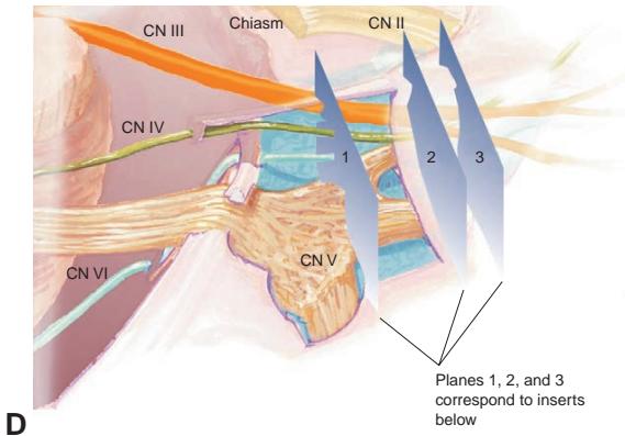


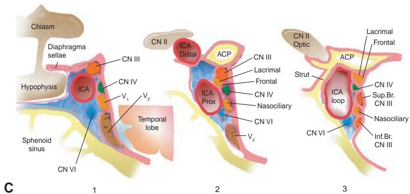
Figure 1-39 Anatomical relationships of the ocular motor nerves. A, Subarachnoid course of the ocular motor nerves. Note their relationship to the surrounding dural structures, particularly the tentorium (partially removed to visualize CNs) and the dura of the clivus. CN III enters the dural canal at the posterior aspect of the cavernous sinus, CN IV enters at the tentorial edge, and CN VI enters along the clivus. B, Major blood vessels and their relationships to the ocular motor nerves. Note the passage of CN III between the superior cerebellar artery below and the posterior cere bral artery above. CN VI runs by the anterior inferior cerebellar artery, which is a major branch off the basilar artery. C, Intracavernous course of the ocular motor nerves. CNs III and IV run in the lateral wall of the cavernous sinus along with CN V divisions $ V_{1} $ and $ V_{2} $. CN VI runs close to the carotid artery within the cavernous sinus itself. D, As the nerves course toward the anterior aspect of the cavernous sinus and the superior orbital fissure, the ophthalmic branch of CN V (CN $ V_{1} $) divides into 3 branches: the lacrimal, frontal, and nasociliary nerves. ACoA = anterior communi cating artery; ACP = anterior clinoid process; ICA = internal carotid artery; Inf. Br. = inferior branch; Prox = proximal; Sup. Br. = superior branch. (Illustrations by Craig A. Luce.)
Extraocular Muscles
The final common pathways that influence the position of the eye within the orbit are the numerous soft-tissue elements connected to the globe. In addition to the EOMs, these tis sues include the optic nerve, Tenon capsule, blood vessels, and the conjunctiva anteriorly. (For a discussion of orbital anatomy, see BCSC Section 7, Oculofacial Plastic and Orbital Surgery.)
Of the 6 EOMs, 4 are rectus muscles (lateral, medial, superior, and inferior), and 2 are oblique muscles (superior and inferior). The rectus muscles originate—along with the levator palpebrae superioris muscle—at the annulus of Zinn, a condensation of tis sue around the optic nerve at the orbital apex. They run forward within sheaths that are connected by intermuscular septa to pierce the posterior Tenon capsule and insert on the anterior sclera at points variably posterior to the corneal limbus, increasing from the me dial through the inferior and lateral to the superior insertion (spiral of Tillaux). The rectus muscles are also held in position by septal attachments to the orbital periosteum, which act as pulleys.
The 2 oblique muscles insert on the posterior lateral aspect of the globe. The inferior oblique muscle originates in the anteromedial periorbita near the posterior margin of the lacrimal fossa. The effective origin of the superior oblique muscle is the trochlea, a pulley- like structure located at the notch in the superior medial orbit. The superior oblique muscle runs anteriorly in the superior medial orbit to the trochlea, where its tendon’s direction of action is reversed.
The EOMs have variable mass and cross section. The inferior oblique is the thinnest, and the medial rectus is the largest. Thus, with normal tonic innervation, the somewhat stronger medial rectus reduces divergent phoria. With the exception of the medial and lateral rectus muscles, the EOMs have primary, secondary, and tertiary functions that vary depending on the position of gaze (Fig 1-40). For further discussion and illustration of the EOMs and their actions, see BCSC Section 6, Pediatric Ophthalmology and Strabismus. Also see Chapter 8 for the clinical etiologies of EOM dysfunction.
Sensory and Facial Motor Anatomy
Cranial nerves V and VII contribute significantly to normal ophthalmic function and are frequently involved in neuro-ophthalmic disorders. Proper functioning of CN V pre serves corneal sensation, loss of which may be accompanied by abnormal corneal epithe lial growth (neurotrophic keratitis associated with loss of neural secreted growth factors). A functioning CN VII prevents exposure keratopathy. Dysfunction of both CNs V and VII greatly increases the risk of corneal damage.