КЛИНИЧЕСКИЙ СОВЕТ
CLINICAL PEARL
Поражения ската (clivus) могут вызвать односторонний или двусторонний паралич CN VI.
Lesions of the clivus can cause unilateral or bilateral CN VI palsies.
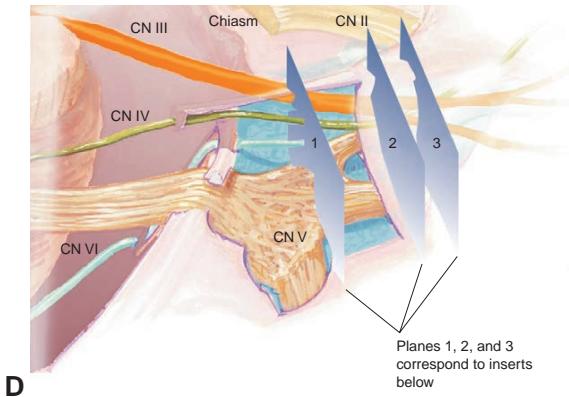


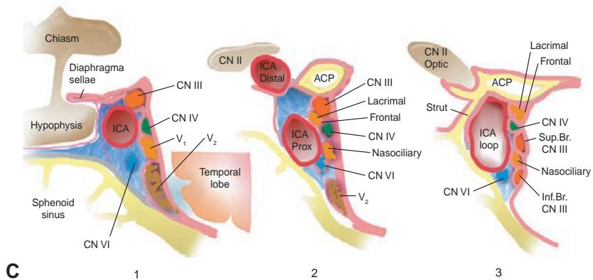
Рисунок 1-39 Анатомические взаимоотношения глазодвигательных нервов. A, Субарахноидальный ход глазодвигательных нервов. Обратите внимание на их взаимоотношение с окружающими структурами твердой мозговой оболочки, в частности с наметом мозжечка (частично удален для визуализации ЧН) и твердой мозговой оболочкой ската. CN III входит в дуральный канал с задней стороны кавернозного синуса, CN IV входит по краю намета, а CN VI входит вдоль ската. B, Основные кровеносные сосуды и их взаимоотношения с глазодвигательными нервами. Обратите внимание на прохождение CN III между верхней мозжечковой артерией снизу и задней мозговой артерией сверху. CN VI проходит рядом с передней нижней мозжечковой артерией, которая является крупной ветвью базилярной артерии. C, Интракавернозный ход глазодвигательных нервов. CN III и IV проходят в латеральной стенке кавернозного синуса вместе с ветвями CN V $ V_{1} $ и $ V_{2} $. CN VI проходит рядом с сонной артерией непосредственно внутри кавернозного синуса. D, As the nerves course toward the anterior aspect of the cavernous sinus and the superior orbital fissure, the ophthalmic branch of CN V (CN $ V_{1} $) делится на 3 ветви: слезный, лобный и носоресничные нервы. ACoA = передняя соединительная артерия; ACP = передний клиноидный отросток; ICA = внутренняя сонная артерия; Inf. Br. = нижняя ветвь; Prox = проксимальный; Sup. Br. = верхняя ветвь. (Иллюстрации Craig A. Luce.)
Figure 1-39 Anatomical relationships of the ocular motor nerves. A, Subarachnoid course of the ocular motor nerves. Note their relationship to the surrounding dural structures, particularly the tentorium (partially removed to visualize CNs) and the dura of the clivus. CN III enters the dural canal at the posterior aspect of the cavernous sinus, CN IV enters at the tentorial edge, and CN VI enters along the clivus. B, Major blood vessels and their relationships to the ocular motor nerves. Note the passage of CN III between the superior cerebellar artery below and the posterior cere bral artery above. CN VI runs by the anterior inferior cerebellar artery, which is a major branch off the basilar artery. C, Intracavernous course of the ocular motor nerves. CNs III and IV run in the lateral wall of the cavernous sinus along with CN V divisions $ V_{1} $ and $ V_{2} $. CN VI runs close to the carotid artery within the cavernous sinus itself. D, As the nerves course toward the anterior aspect of the cavernous sinus and the superior orbital fissure, the ophthalmic branch of CN V (CN $ V_{1} $) divides into 3 branches: the lacrimal, frontal, and nasociliary nerves. ACoA = anterior communi cating artery; ACP = anterior clinoid process; ICA = internal carotid artery; Inf. Br. = inferior branch; Prox = proximal; Sup. Br. = superior branch. (Illustrations by Craig A. Luce.)
Глазодвигательные мышцы
Extraocular Muscles
Конечными общими путями, влияющими на положение глаза в орбите, являются многочисленные мягкотканные элементы, соединенные с глазным яблоком. Помимо глазодвигательных мышц, эти ткани включают зрительный нерв, капсулу Тенона, кровеносные сосуды и конъюнктиву спереди. (Для обсуждения анатомии орбиты см. BCSC Раздел 7, Oculofacial Plastic and Orbital Surgery.)
The final common pathways that influence the position of the eye within the orbit are the numerous soft-tissue elements connected to the globe. In addition to the EOMs, these tis sues include the optic nerve, Tenon capsule, blood vessels, and the conjunctiva anteriorly. (For a discussion of orbital anatomy, see BCSC Section 7, Oculofacial Plastic and Orbital Surgery.)
Из 6 глазодвигательных мышц 4 являются прямыми мышцами (наружной, внутренней, верхней и нижней) и 2 — косыми мышцами (верхней и нижней). Прямые мышцы берут начало — вместе с верхней мышцей, поднимающей верхнее веко — от кольца Цинна, уплотнения ткани вокруг зрительного нерва у верхушки орбиты. Они идут вперед в оболочках, которые соединены межмышечными перегородками, чтобы пронзить заднюю капсулу Тенона и прикрепиться к передней склере в точках, расположенных с разным расстоянием позади лимба роговицы, увеличивающемся от медиального через нижний и латеральный к верхнему прикреплению (спираль Тильо). Прямые мышцы также удерживаются в положении за счет перегородочных прикреплений к надкостнице орбиты, которые действуют как блоки.
Of the 6 EOMs, 4 are rectus muscles (lateral, medial, superior, and inferior), and 2 are oblique muscles (superior and inferior). The rectus muscles originate—along with the levator palpebrae superioris muscle—at the annulus of Zinn, a condensation of tis sue around the optic nerve at the orbital apex. They run forward within sheaths that are connected by intermuscular septa to pierce the posterior Tenon capsule and insert on the anterior sclera at points variably posterior to the corneal limbus, increasing from the me dial through the inferior and lateral to the superior insertion (spiral of Tillaux). The rectus muscles are also held in position by septal attachments to the orbital periosteum, which act as pulleys.
2 косые мышцы прикрепляются к заднелатеральной поверхности глазного яблока. Нижняя косая мышца берет начало в переднемедиальной периорбите рядом с задним краем слезной ямки. Эффективным началом верхней косой мышцы является трохлея — блокподобная структура, расположенная в вырезке в верхнемедиальной части орбиты. Верхняя косая мышца идет вперед в верхнемедиальной части орбиты к трохлее, где направление действия ее сухожилия меняется на противоположное.
The 2 oblique muscles insert on the posterior lateral aspect of the globe. The inferior oblique muscle originates in the anteromedial periorbita near the posterior margin of the lacrimal fossa. The effective origin of the superior oblique muscle is the trochlea, a pulley- like structure located at the notch in the superior medial orbit. The superior oblique muscle runs anteriorly in the superior medial orbit to the trochlea, where its tendon’s direction of action is reversed.
Глазодвигательные мышцы имеют различную массу и площадь поперечного сечения. Нижняя косая мышца является самой тонкой, а внутренняя прямая мышца — самой крупной. Таким образом, при нормальной тонической иннервации несколько более сильная внутренняя прямая мышца уменьшает дивергентную форию. За исключением внутренней и наружной прямых мышц, глазодвигательные мышцы имеют первичные, вторичные и третичные функции, которые варьируются в зависимости от положения взора (рис. 1-40). Для дальнейшего обсуждения и иллюстрации глазодвигательных мышц и их действий см. BCSC Раздел 6, Pediatric Ophthalmology and Strabismus. Также см. Главу 8 для ознакомления с клиническими этиологиями дисфункции глазодвигательных мышц.
The EOMs have variable mass and cross section. The inferior oblique is the thinnest, and the medial rectus is the largest. Thus, with normal tonic innervation, the somewhat stronger medial rectus reduces divergent phoria. With the exception of the medial and lateral rectus muscles, the EOMs have primary, secondary, and tertiary functions that vary depending on the position of gaze (Fig 1-40). For further discussion and illustration of the EOMs and their actions, see BCSC Section 6, Pediatric Ophthalmology and Strabismus. Also see Chapter 8 for the clinical etiologies of EOM dysfunction.
Сенсорная и лицевая моторная анатомия
Sensory and Facial Motor Anatomy
Черепные нервы V и VII вносят значительный вклад в нормальную функцию глаза и часто вовлечены в нейроофтальмологические расстройства. Надлежащее функционирование CN V сохраняет чувствительность роговицы, утрата которой может сопровождаться аномальным ростом эпителия роговицы (нейротрофический кератит, связанный с потерей секретируемых нейронами факторов роста). Функционирующий CN VII предотвращает экспозиционную кератопатию. Дисфункция обоих ЧН, V и VII, значительно увеличивает риск повреждения роговицы.
Cranial nerves V and VII contribute significantly to normal ophthalmic function and are frequently involved in neuro-ophthalmic disorders. Proper functioning of CN V pre serves corneal sensation, loss of which may be accompanied by abnormal corneal epithe lial growth (neurotrophic keratitis associated with loss of neural secreted growth factors). A functioning CN VII prevents exposure keratopathy. Dysfunction of both CNs V and VII greatly increases the risk of corneal damage.