Vestibular ocular system (vestibular ocular reflex)
The vestibular nuclei provide major supranuclear input into ocular motility and tonic input into eye positioning.
The vestibular nerve and cochlear nerve form the CN VIII complex and exit the petrous bone through the internal auditory meatus.
CN VIII traverses the subarachnoid space within the cerebellopontine angle to me dial, lateral, and superior vestibular nuclei in the medulla.
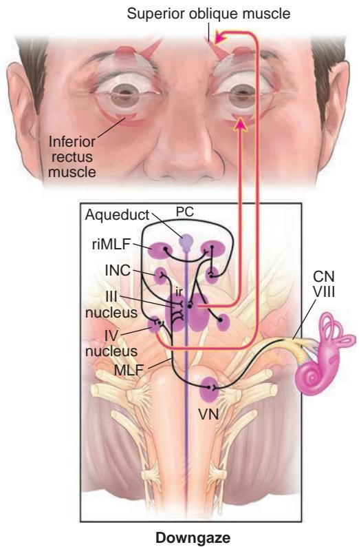

Figure 1-32 Anatomical schemes for the synthesis of vertical eye movements. From the vertical semicircular canals, primary afferents from CN VIII synapse on the vestibular nuclei (VN) and ascend into the medial longitudinal fasciculus (MLF) and brachium conjunctivum (not shown) to the CN III and CN IV nuclei and the interstitial nucleus of Cajal (INC). Excitatory burst neurons in the rostral interstitial nucleus of the MLF (riMLF) project to the motoneurons of CNs III and IV and send axon collaterals to the INC. III nucleus = oculomotor nerve (CN III) nucleus; IV nucleus = trochlear nerve (CN IV) nucleus; PC = posterior commissure. (Illustration by Rob Flewell, CMI.)
The medial and inferior vestibular nuclei project to the nodulus (a central nucleus of the cerebellum) and the ventral uvula.
For information on clinical disorders of vestibular-ocular function, see Chapter 9.
Ocular Motor Cranial Nerves
Without neural activity, the visual axes are usually mildly to moderately divergent. The major tonic input to ocular motility is supplied by 3 pairs of ocular motor CNs—III, IV, and VI—that innervate the 6 EOMs controlling ocular movement (Fig 1-34). In addition, CN III innervates the levator palpebrae superioris and the pupillary sphincter muscles.
Except for the inferior oblique muscle, the innervation of the EOMs occurs at ap proximately one-third of the distance from the apex. The inferior oblique muscle receives innervation at approximately its midpoint from a neurovascular bundle running parallel to the lateral aspect of the inferior rectus muscle. Except for the superior oblique muscle, all EOMs receive their innervation on the inside surface. The branches of CN IV termi nate on the upper (outer) surface of the muscle.

Figure 1-33 Anatomical scheme for the synthesis of horizontal eye movements. From the hori zontal semicircular canal, primary afferents of the vestibular nerve (CN VIII) project mainly to neurons of the vestibular nerve nucleus (VIII nucleus), which then send an excitatory connection to the contralateral CN VI nucleus. CN VI innervates the ipsilateral lateral rectus muscle and the contralateral CN III nucleus via the medial longitudinal fasciculus (MLF). Horizontal saccades are generated in the frontal eye field, which activates the contralateral paramedian pontine reticu lar formation (PPRF). Burst neurons in the PPRF stimulate the ipsilateral CN VI nucleus, with the subsequent pathway identical to that subserving horizontal vestibular-generated eye move ments. Some neurons in the PPRF project to the nucleus prepositus hypoglossi-medial vestibu lar nucleus (neural integrator) and then to the CN VI nucleus. (Illustration by Rob Flewell, CMI.)
See Chapter 8 for information on the clinical presentation of disorders due to infra- nuclear, fascicular, and peripheral ocular motor CN lesions.
Third cranial nerve
The CN III (oculomotor) nucleus is located dorsally within the midbrain ventral to the aqueduct connecting the third and fourth ventricles (Fig 1-35). The nuclear complex itself represents a collection of subnuclei that have specific identifiable functions (Fig 1-36). The fibers destined to innervate the medial rectus, inferior rectus, inferior oblique, pupil sphincter, and ciliary body muscles exit ventrally ipsilateral to the individual nuclei from which they originate. In contrast, the fibers from the medial subnucleus, which lies along the midline, cross before exiting the brainstem to innervate the contralateral superior rec tus muscle. The central caudate nucleus provides innervation to the levator palpebrae superioris bilaterally. A nuclear CN III lesion can cause an ipsilateral CN III palsy with possible additional limitation of supraduction of the contralateral eye and/or bilateral pto sis depending on which subnuclei are affected.
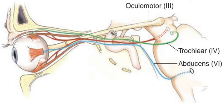
Figure 1-34 Lateral view of the course of CNs III, IV, and VI. (Illustration by Dave Peace.)
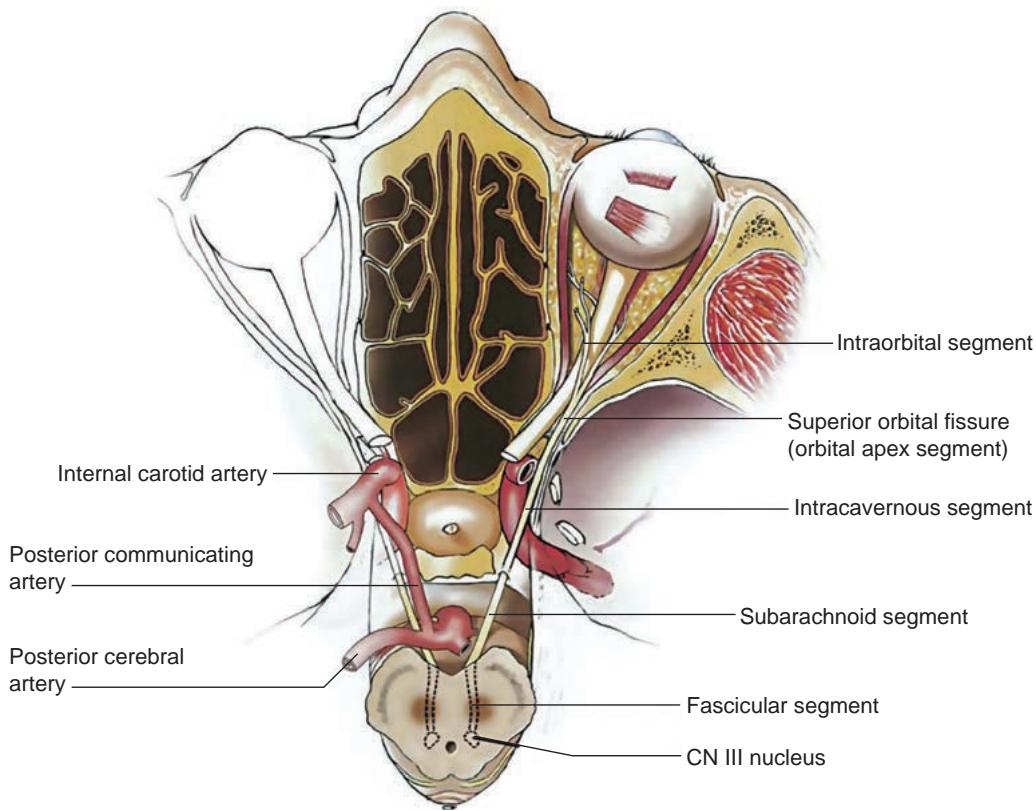
Figure 1-35 Illustration of the axial course of CN III from its origin in the midbrain. (Reprinted from Foroozan R, Bhatti MT, Rhoton AL. Transsphenoidal diplopia. Surv Ophthalmol. 2004;49(3):352; with permission from Elsevier.)