The parastriate cortex (also called V2, or Brodmann area 18) is contiguous with the pri mary visual cortex and receives its input from V1. Area V3 projects primarily to the posterior parietal lobe and receives direct input from V1. Area V3 has no sharp histologic delineation from V2 and sends efferent information to the basal ganglia (pulvinar) and the midbrain. Cells in this area are thought to respond to more than 1 stimulus dimension, suggesting that visual integration occurs in this region. Area V4, located within the lingual and fusiform gyri, is particularly sensitive to color. Damage to this area is probably responsible for most cases of cerebral achromatopsia. Area V5 is located posterior to and within the superior temporal sulcus and gyrus subangularis. It corresponds to the middle temporal visual region and re ceives ipsilateral input from V1 and direct input from the M-cell layers of the LGN (Fig 1-27). The receptive fields of V5 are larger than those of V1, and the underlying white matter is heavily myelinated. The SC receives afferent input directly from the anterior visual pathways and indirectly from the calcarine cortex. The superficial layers contain a retinotopic map that overlies the deeper layers, which are primarily concerned with saccadic generation.
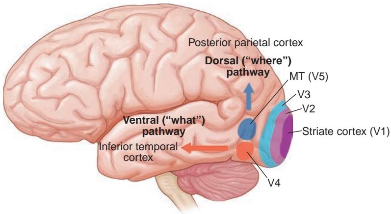
Figure 1-27 Parallel visual processing pathways in the human brain. The occipitotemporal (ven tral, or “what”) pathway begins in the striate cortex (V1) and projects to the angular gyrus for language processing, to the inferior temporal cortex for object identification, and to the limbic structures. The occipitoparietal (dorsal, or “where”) pathway begins in the striate cortex and projects to the posterior parietal cortex and the superior temporal cortex and is responsible for visual–spatial analysis. MT = middle temporal visual area.
CLINICAL PEARL
The neurons in area V5 encode the speed and direction of moving stimuli. This sensory area is likely the origin of pursuit movements and thus connects the afferent and efferent pathways.
Horton JC, Hoyt WF. The representation of the visual field in human striate cortex.
A revision of the classic Holmes map. Arch Ophthalmol. 1991;109(6):816–824.
Efferent Visual System
Over the last few decades, our understanding of the anatomy, physiology, and pathology of the ocular motor system has increased dramatically on the basis of primate model experi ments, human electrophysiology testing, functional magnetic resonance imaging studies, and the clinical-pathologic-radiologic correlations of disorders in patients with eye movement abnormalities.
A properly functioning ocular motor system ultimately ensures clear, stable, and bin ocular vision. Two basic human eye movements facilitate this task:
gaze shift
gaze stabilization
These movements can be further divided into 6 functional systems:
ocular fixation system
vestibular ocular system (vestibular ocular reflex)
optokinetic system
saccadic system
smooth-pursuit system
vergence system
Although these systems are under the control of—and are modulated by—different re gions of the brain (ie, the cortex) and brainstem, they have considerable anatomical and functional overlaps (see Chapter 9).
This section provides an overview of the anatomy of the ocular motor system, with a detailed discussion of clinical applications presented in Chapter 9. To facilitate learning, this discussion follows a top-to-bottom approach based on the following topics:
cortical control of eye movements, including frontal eye fields, supplementary eye fields, and posterior parietal cortex
subcortical centers, including the basal ganglia (BG), thalamus, and SC
brainstem or premotor structures coordinating conjugate eye movements, includ ing the vestibular-ocular system and the cerebellum
ocular motor CNs (CNs III, IV, and VI)
extraocular muscles (EOMs)
Interested readers can find a comprehensive description of the ocular motor system in the following reference.
Leigh RJ, Zee DS. The Neurology of Eye Movements. 5th ed. Contemporary Neurology Series.
Oxford University Press; 2015.
Cortical Input
The efferent visual system spans a large segment of the central nervous system, as many areas of the brain generate eye movements (Fig 1-28). Saccadic and smooth-pursuit eye movements were once thought to derive from distinct supranuclear pathways; however, it now appears that these systems overlap considerably. In addition, there are 2 major visual pathways: one for the movement of images (magnocellular: M cells) and the other for the discrimination of images (parvocellular: P cells).
Saccadic system
The saccadic system comprises the following structures in an organized framework (Fig 1-29):
cortical structures for volitional/memory-guided saccades: frontal eye fields, sup plementary eye fields; visually reflexive saccades, which are generated by various areas of the parietal cortex but mainly from the superior parietal lobe (Brodmann area 7) and parietal eye fields with connections to the SC and frontal and supple mentary eye fields
subcortical structures: the BG, thalamus, and SC
brainstem neural network or premotor neurons: several types of pontine neurons
brainstem saccade generators: PPRF and rostral interstitial nucleus of the medial longitudinal fasciculus (riMLF)
motoneurons of the ocular motor CNs
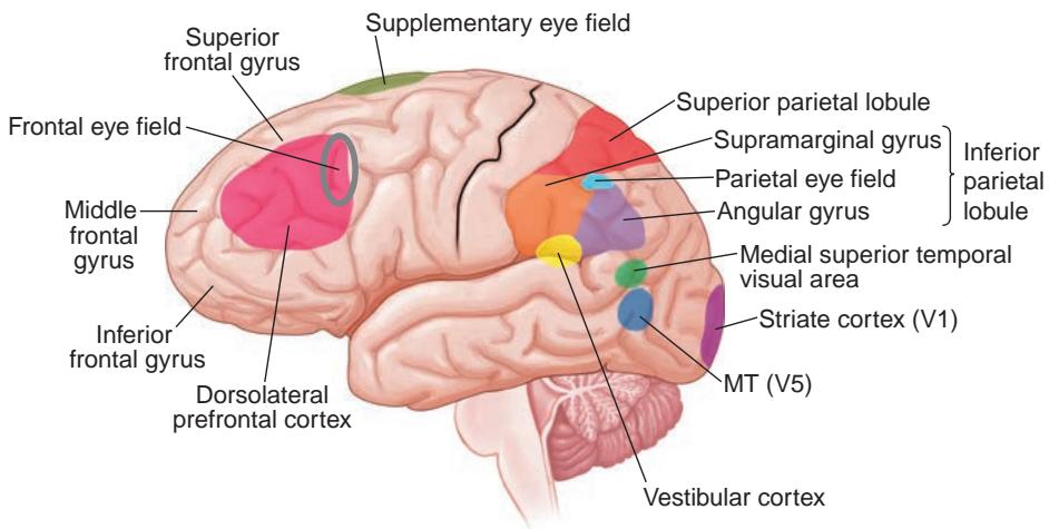
Figure 1-28 Overview of the cortical centers that control human eye movements. MT = middle temporal visual area. (Redrawn by Rob Flewell, CMI.)
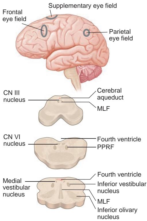
Figure 1-29 The saccadic system comprises visually reflexive movements (derived from the parietal eye field) and memory-guided and voli tional movements (derived from the frontal eye field). Supplemental eye fields receive input from frontal eye fields and program saccades projecting to the superior colliculus, basal gan glia, and thalamus. Projections are also sent to the paramedian pontine reticular formation (PPRF); rostral interstitial medial longitudinal fasciculus, which is more rostral in the mid brain; and motoneurons of the ocular motor cranial nerves (CNs). MLF = medial longitudinal fasciculus. (Illustration by Cyndie C. H. Wooley.)