Зрительный тракт
Optic Tract
Волокна, выходящие из зрительного перекреста, проходят по окружности вокруг промежуточного мозга латеральнее гипоталамуса и контактируют с амбиентной цистерной (см. рис. 1-13). Отсутствие близости между соответствующими волокнами от правого и левого глаз объясняет неконгруэнтный характер дефектов поля зрения при поражении зрительного тракта. Непосредственно перед LGN волокна, участвующие в пупиллярных путях, отходят к претектальным ядрам; другие волокна отходят к поверхностным слоям верхних холмиков (SC) через brachium SC. Эти волокна, которые берут начало в ipRGC, вероятно, являются единственным источником пупилломоторного сигнала от сетчатки к среднему мозгу. ipRGC также проецируются на супрахиазматическое ядро гипоталамуса, которое, вероятно, отвечает за индуцированные светом суточные ритмы. Большинство аксонов, берущих начало в RGC, заканчиваются в LGN.
The fibers exiting the optic chiasm proceed circumferentially around the diencephalon lateral to the hypothalamus and in contact with the ambient cistern (see Fig 1-13). The lack of proximity between corresponding fibers from the right and left eyes explains the incongruous nature of optic tract visual field defects. Just before the LGN, the fibers in volved in the pupillary pathways exit to the pretectal nuclei; other fibers exit to the super ficial layers of the superior colliculi (SC) via the brachium of the SC. These fibers, which originate from ipRGCs, are likely the sole source of pupillomotor input from the retina to the midbrain. The ipRGCs also project to the suprachiasmatic nucleus of the hypothala mus, which is likely responsible for light-induced diurnal rhythms. Most of the axons that originate in the RGCs terminate within the LGN.
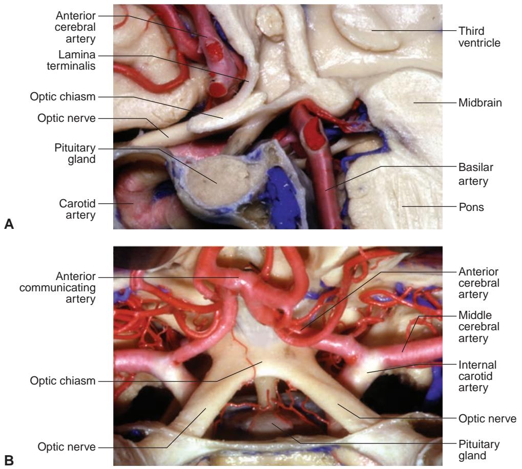
Рисунок 1-23 Анатомическая диссекция зрительного перекреста и окружающих структур. A, Сагиттальный вид. B, Верхний вид. (Предоставлено Albert L. Rhoton Jr, MD.)
Figure 1-23 Anatomical dissection of the optic chiasm and surrounding structures. A, Sagittal view. B, Superior view. (Courtesy of Albert L. Rhoton Jr, MD.)
LGN располагается в задней части таламуса ниже и латеральнее пульвинара и выше латерального рецессуса амбиентной цистерны. Эта остроконечная структура грибовидной формы разделена на 6 уровней (см. рисунок 3-9 в BCSC Section 2, Fundamentals and Principles of Ophthalmology): аксоны, берущие начало в контралатеральном глазу, заканчиваются в 1, 4 и 6 слоях LGN. Аксоны из ипсилатерального глаза заканчиваются во 2, 3 и 5 слоях.
The LGN is located in the posterior thalamus below and lateral to the pulvinar and above the lateral recess of the ambient cistern. This peaked, mushroom-shaped structure is divided into 6 levels (see Figure 3-9 in BCSC Section 2, Fundamentals and Principles of Ophthalmology): Axons originating in the contralateral eye terminate in layers 1, 4, and 6 of the LGN. Axons from the ipsilateral eye terminate in layers 2, 3, and 5.
Два нижних слоя (слои 1 и 2) получают сигнал от волокон M-клеток — ганглионарных клеток с более крупными рецептивными полями, чувствительными к обнаружению движения.
Two inferior layers (layers 1 and 2) receive input from fibers of M-cells, ganglion cells with larger receptive fields sensitive to motion detection.
Четыре верхних уровня (слои 3, 4, 5 и 6) являются терминалями аксонов P-клеток — ганглионарных клеток с более мелкими рецептивными полями, отвечающими за обеспечение максимального пространственного разрешения и цветовосприятия.
Four superior levels (layers 3, 4, 5, and 6) are the termini of axons of P-cells, gan glion cells with smaller receptive fields responsible for mediating maximal spatial resolution and color perception.
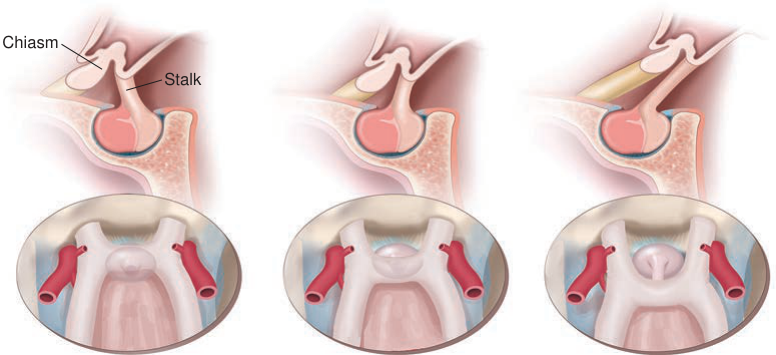
Рисунок 1-24 Положение зрительного перекреста по отношению к турецкому седлу. (Иллюстрация Rob Flewell, CMI.)
Figure 1-24 Position of the optic chiasm in relation to the sella. (Illustration by Rob Flewell, CMI.)
Префиксный
Prefixed
Нормальный
Normal
Постфиксный
Postfixed
(нормальная конфигурация: 85%)
(normal configuration: 85%)
По мере приближения волокон к LGN ретинальная репрезентация поворачивается почти 90°, при этом верхние волокна смещаются медиально, а нижние — латерально. Макулярные волокна имеют тенденцию смещаться суперлатерально. Корковые и субкорковые пути могут модулировать активность в LGN. Кроме того, кора, SC и претектальные ядра проецируются обратно в LGN.
As the fibers approach the LGN, the retinal representation rotates almost 90°, with the superior fibers moving medially and the inferior fibers laterally. The macular fibers tend to move superolaterally. Cortical and subcortical pathways may modulate activity in the LGN. In addition, the cortex, SC, and pretectal nuclei project back to the LGN.
Кора
Cortex
После синапса в LGN аксоны направляются кзади в виде зрительных лучистостей, которые заканчиваются в первичной зрительной (калькариновой) коре в затылочной доле (рис. 1-25). Самые нижние волокна сначала идут вперед, затем латерально и назад, образуя петлю вокруг височного рога боковых желудочков (называемую петлей Мейера). Верхние волокна идут назад через глубокое белое вещество теменной доли. Макулярные (центральные) волокна проходят латерально, в то время как периферические волокна более сконцентрированы в верхних и нижних отделах лучистостей.
Following a synapse in the LGN, axons travel posteriorly as optic radiations that terminate in the primary visual (calcarine) cortex in the occipital lobe (Fig 1-25). The most inferior fibers first travel anteriorly, then laterally and posteriorly to loop around the temporal horn of the lateral ventricles (called the Meyer loop). The superior fibers travel posteriorly through the deep white matter of the parietal lobe. The macular (central) fibers course laterally, with the peripheral fibers concentrated more at the superior and inferior aspects of the radiations.
Повреждение волокон внутри лучистости вызывает гомонимную гемианопсию — контралатеральный дефект поля зрения, который соблюдает вертикальную срединную линию. Если соответствующие волокна от двух глаз расположены близко друг к другу, дефект поля зрения в каждом глазу идентичен, или конгруэнтен. Конгруэнтные дефекты поля зрения возникают в результате поражений в калькариновой коре. Более переднее вовлечение часто вызывает неконгруэнтные дефекты поля зрения, что позволяет предположить, что соответствующие волокна расположены дальше друг от друга более спереди в зрительных путях.
Injury to fibers within a radiation produces a homonymous hemianopia, a contralat eral visual field defect that respects the vertical midline. If the corresponding fibers from the 2 eyes are close to each other, the visual field defect in each eye is identical, or con gruous. Congruous visual field defects occur as a result of lesions in the calcarine cortex. More anterior involvement often produces incongruous visual field defects, suggesting that the corresponding fibers lie farther apart more anteriorly in the visual pathways.
Первичная зрительная кора (известная также как V1, полосатая кора или поле Бродмана 17) расположена вдоль горизонтальной калькариновой щели, которая разделяет медиальную поверхность затылочной доли. Волокна зрительных лучистостей заканчиваются в четвертом из 6 слоев первичной зрительной коры. Волокна из височного полумесяца, или самой латеральной части поля зрения (берущие начало только в контралатеральном глазу), заканчиваются наиболее спереди (рис. 1-26). Макулярные волокна заканчиваются более кзади.
The primary visual cortex (known variously as V1, striate cortex, or Brodmann area 17) is arrayed along the horizontal calcarine fissure, which divides the medial surface of the occipital lobe. Fibers of the optic radiations terminate in the fourth of 6 layers in the primary visual cortex. Fibers from the temporal crescent, or the most lateral visual field (originating only in the contralateral eye), terminate most anteriorly (Fig 1-26). The macular fibers terminate more posteriorly.

Рисунок 1-25 Схема зрительных лучистостей, сагиттальный вид. Нижние волокна (обеспечивающие верхнее поле зрения) идут вперед, прежде чем повернуть назад в височной доле. Верхние волокна (обеспечивающие нижнее поле зрения) идут дорсально в теменной доле, чтобы закончиться в затылочной доле выше калькариновой щели. (Перерисовано с разрешения University of Texas at Dallas. Иллюстрация Mark Miller.)
Figure 1-25 Schematic of the optic radiations, sagittal view. The inferior fibers (subserving the superior visual field) course anteriorly before looping posteriorly in the temporal lobe. The superior fibers (subserving the inferior visual field) course dorsally in the parietal lobe to terminate in the occipital lobe above the calcarine fissure. (Redrawn with permission from the University of Texas at Dallas. Illustration by Mark Miller.)
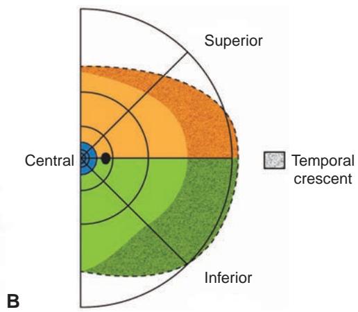
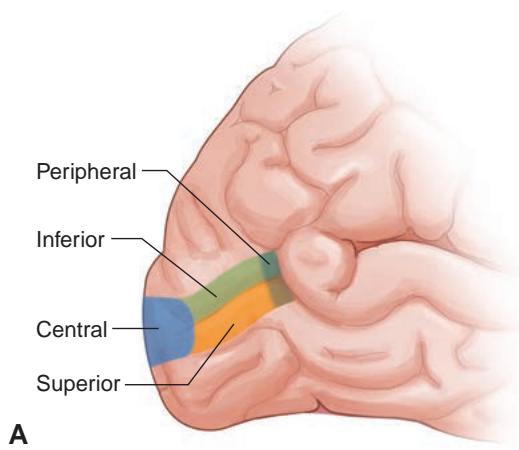
Рисунок 1-26 Первичная зрительная кора и соответствующая репрезентация поля зрения. A, Левая затылочная кора, показывающая расположение полосатой коры внутри калькариновой щели. Синим цветом обозначена макула (центральное поле зрения); зеленым — нижнее поле зрения; оранжевым — верхнее поле зрения. Самые периферические волокна представлены пунктирными цветами. B, Правая половина поля зрения соответствует областям полосатой коры на рис. A. Пунктирная область соответствует монокулярному височному полумесяцу, который картируется в самых передних примерно 8% полосатой коры. Черная точка = физиологическое слепое пятно. (Иллюстрации Christine Gralapp.)
Figure 1-26 Primary visual cortex and corresponding visual field representation. A, Left oc cipital cortex showing the location of the striate cortex within the calcarine fissure. Blue rep resents the macula (central visual field); green represents the inferior visual field; and orange represents the superior visual field. The most peripheral fibers are represented by the stippled colors. B, Right hemifield corresponds to the regions of the striate cortex in A. The stippled area corresponds to the monocular temporal crescent, which is mapped in the most anterior approxi mately 8% of the striate cortex. Black dot = physiologic blind spot. (Illustrations by Christine Gralapp.)
Кора в значительной степени ориентирована на центральную ретинальную активность: 50%–60% коры реагируют на активность в пределах центральных 10° и примерно 80% посвящено макулярной активности (в пределах 30°).
The cortex is heavily weighted to central retinal activity, with 50%–60% of the cortex responding to activity within the central 10° and approximately 80% devoted to macular activity (within 30°).
Верхняя часть коры продолжает получать информацию из нижнего поля зрения в ретинотопическом распределении. Такое ретинотопическое картирование на всем протяжении афферентных зрительных путей позволяет локализовать поражения на основании дефектов поля зрения. Антеромедиальная часть полосатой коры представляет дальнее монокулярное височное поле зрения (височный полумесяц) контралатерального глаза. Таким образом, дефект дальнего монокулярного височного поля зрения локализует поражение в контралатеральной передней затылочной коре (см. рис. 1-26).
The superior portion of the cortex continues to receive information from the inferior visual field in a retinotopic distribution. This retinotopic mapping throughout the afferent visual pathways allows lesions to be localized on the basis of visual field defects. The an teromedial portion of the striate cortex represents the far monocular temporal visual field (temporal crescent) of the contralateral eye. Therefore, a far monocular temporal visual field defect localizes the lesion to the contralateral anterior occipital cortex (see Fig 1-26).