Optic Tract
The fibers exiting the optic chiasm proceed circumferentially around the diencephalon lateral to the hypothalamus and in contact with the ambient cistern (see Fig 1-13). The lack of proximity between corresponding fibers from the right and left eyes explains the incongruous nature of optic tract visual field defects. Just before the LGN, the fibers in volved in the pupillary pathways exit to the pretectal nuclei; other fibers exit to the super ficial layers of the superior colliculi (SC) via the brachium of the SC. These fibers, which originate from ipRGCs, are likely the sole source of pupillomotor input from the retina to the midbrain. The ipRGCs also project to the suprachiasmatic nucleus of the hypothala mus, which is likely responsible for light-induced diurnal rhythms. Most of the axons that originate in the RGCs terminate within the LGN.
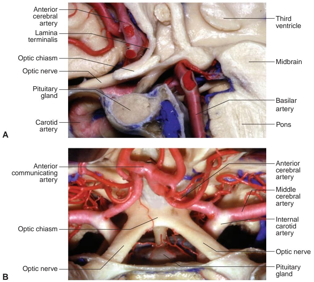
Figure 1-23 Anatomical dissection of the optic chiasm and surrounding structures. A, Sagittal view. B, Superior view. (Courtesy of Albert L. Rhoton Jr, MD.)
The LGN is located in the posterior thalamus below and lateral to the pulvinar and above the lateral recess of the ambient cistern. This peaked, mushroom-shaped structure is divided into 6 levels (see Figure 3-9 in BCSC Section 2, Fundamentals and Principles of Ophthalmology): Axons originating in the contralateral eye terminate in layers 1, 4, and 6 of the LGN. Axons from the ipsilateral eye terminate in layers 2, 3, and 5.
Two inferior layers (layers 1 and 2) receive input from fibers of M-cells, ganglion cells with larger receptive fields sensitive to motion detection.
Four superior levels (layers 3, 4, 5, and 6) are the termini of axons of P-cells, gan glion cells with smaller receptive fields responsible for mediating maximal spatial resolution and color perception.
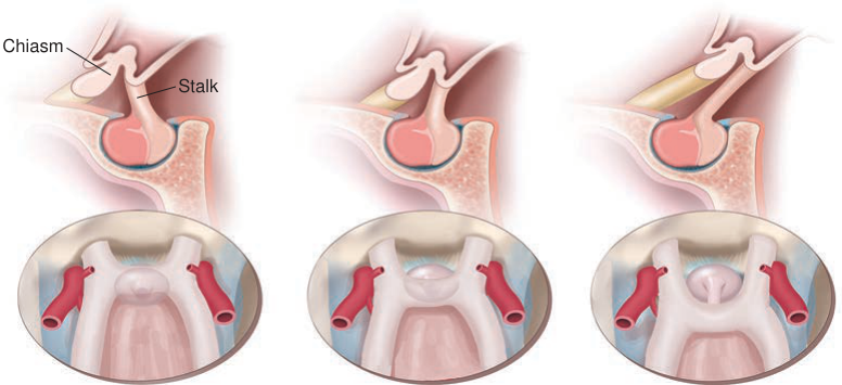
Figure 1-24 Position of the optic chiasm in relation to the sella. (Illustration by Rob Flewell, CMI.)
Prefixed
Normal
Postfixed
(normal configuration: 85%)
As the fibers approach the LGN, the retinal representation rotates almost 90°, with the superior fibers moving medially and the inferior fibers laterally. The macular fibers tend to move superolaterally. Cortical and subcortical pathways may modulate activity in the LGN. In addition, the cortex, SC, and pretectal nuclei project back to the LGN.
Cortex
Following a synapse in the LGN, axons travel posteriorly as optic radiations that terminate in the primary visual (calcarine) cortex in the occipital lobe (Fig 1-25). The most inferior fibers first travel anteriorly, then laterally and posteriorly to loop around the temporal horn of the lateral ventricles (called the Meyer loop). The superior fibers travel posteriorly through the deep white matter of the parietal lobe. The macular (central) fibers course laterally, with the peripheral fibers concentrated more at the superior and inferior aspects of the radiations.
Injury to fibers within a radiation produces a homonymous hemianopia, a contralat eral visual field defect that respects the vertical midline. If the corresponding fibers from the 2 eyes are close to each other, the visual field defect in each eye is identical, or con gruous. Congruous visual field defects occur as a result of lesions in the calcarine cortex. More anterior involvement often produces incongruous visual field defects, suggesting that the corresponding fibers lie farther apart more anteriorly in the visual pathways.
The primary visual cortex (known variously as V1, striate cortex, or Brodmann area 17) is arrayed along the horizontal calcarine fissure, which divides the medial surface of the occipital lobe. Fibers of the optic radiations terminate in the fourth of 6 layers in the primary visual cortex. Fibers from the temporal crescent, or the most lateral visual field (originating only in the contralateral eye), terminate most anteriorly (Fig 1-26). The macular fibers terminate more posteriorly.

Figure 1-25 Schematic of the optic radiations, sagittal view. The inferior fibers (subserving the superior visual field) course anteriorly before looping posteriorly in the temporal lobe. The superior fibers (subserving the inferior visual field) course dorsally in the parietal lobe to terminate in the occipital lobe above the calcarine fissure. (Redrawn with permission from the University of Texas at Dallas. Illustration by Mark Miller.)
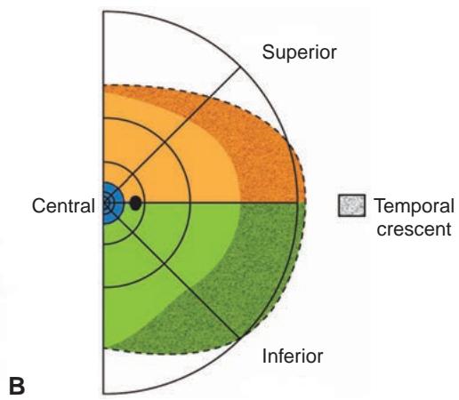
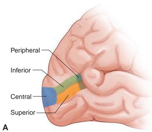
Figure 1-26 Primary visual cortex and corresponding visual field representation. A, Left oc cipital cortex showing the location of the striate cortex within the calcarine fissure. Blue rep resents the macula (central visual field); green represents the inferior visual field; and orange represents the superior visual field. The most peripheral fibers are represented by the stippled colors. B, Right hemifield corresponds to the regions of the striate cortex in A. The stippled area corresponds to the monocular temporal crescent, which is mapped in the most anterior approxi mately 8% of the striate cortex. Black dot = physiologic blind spot. (Illustrations by Christine Gralapp.)
The cortex is heavily weighted to central retinal activity, with 50%–60% of the cortex responding to activity within the central 10° and approximately 80% devoted to macular activity (within 30°).
The superior portion of the cortex continues to receive information from the inferior visual field in a retinotopic distribution. This retinotopic mapping throughout the afferent visual pathways allows lesions to be localized on the basis of visual field defects. The an teromedial portion of the striate cortex represents the far monocular temporal visual field (temporal crescent) of the contralateral eye. Therefore, a far monocular temporal visual field defect localizes the lesion to the contralateral anterior occipital cortex (see Fig 1-26).