

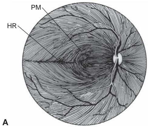
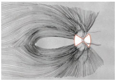
Figure 1-22 Anatomy of retinal ganglion cell axons. A, Pattern of the nerve fiber layer of axons from ganglion cells to the ONH. Superior, inferior, and nasal fibers take a fairly straight course. Temporal axons originate above and below the horizontal raphe (HR) and take an arching course to the ONH. Axons arising from ganglion cells in the nasal macula project directly to the ONH as the papillomacular bundle (PM). B, Lesions involving the decussating nasal retinal fibers (represented by the dashed red line) can result in bow-tie atrophy. C, Schematic depiction of damage to nasal and papillomacular fibers of the retina and patterns of nasal and temporal optic nerve atrophy (represented by red outlined triangles) corresponding to damaged crossing nasal fibers. Therefore, in a patient with a pregeniculate homonymous hemianopia or a bitem poral hemianopia, band (or bow-tie) atrophy occurs with loss of nasal macular and peripheral fibers in the contralateral eye. D, Clinical photograph of a right optic nerve demonstrating bow-tie atrophy. (Part A reprinted from Kline LB, Foroozan R, eds. Optic Nerve Disorders. 2nd ed. Ophthalmology Monographs 10. Oxford University Press, in cooperation with the American Academy of Ophthalmology; 2007:5. Part B illustration by Christine Gralapp; part C courtesy of Neil Miller, MD; part D courtesy of Lanning Kline, MD.)
As the optic nerve enters the optic canal, its dural sheath fuses with the periorbita. It is also surrounded by the annulus of Zinn, which serves as the origin of the 4 rectus muscles and the superior oblique muscle. The optic canal, which runs superiorly and medially, is normally approximately 8–10 mm long and 5–7 mm wide; however, it may be elongated and narrowed by bone thickening processes (eg, fibrous dysplasia, intraosseous meningioma, vitamin A deficiency). Within the canal, the optic nerve is relatively anchored and can easily be injured by shearing forces transmitted from blunt facial trauma (see Chapter 4).
At its intracranial passage, the optic nerve passes under a fold of dura (the falciform ligament) that may impinge on the nerve, especially if it is elevated by lesions arising from the bone of the sphenoid (tuberculum) or the sella. Once it becomes intracranial, the optic nerve no longer has a sheath. The anterior loop of the carotid artery usually lies just below and temporal to the nerve, and the proximal ACA passes over it. The gyrus rectus, the most inferior portion of the frontal lobe, lies above and parallel to the optic nerve. The intracranial portion of the optic nerve—which is 3–16 mm long, usually approximately 10 mm—continues to the optic chiasm.
Lee AG, Morgan ML, Palau A, et al. Anatomy of the optic nerve and visual pathway. In: Tubbs RS, Rizk E, Shoja M, Loukas M, Barbaro N, Spinner R. Nerves and Nerve Injuries. Vol 1. Academic Press; 2015:277–303.
Salazar JJ, Ramirez AI, De Hoz R, et al. Anatomy of the human optic nerve: structure and function. In: Optic Nerve. IntechOpen; 2018. 10.5772/intechopen.79827
Optic Chiasm
The optic chiasm is approximately 12 mm wide, 8 mm long in the anteroposterior di rection, and 4 mm thick (Fig 1-23). It is inclined at almost 45° and is supplied by small arterial branches from the proximal ACA and ACoA. The chiasm is located just anterior to the hypothalamus and the anterior third ventricle (forming part of its anterior wall and causing an invagination) and approximately 10 mm above the sella. Its location with respect to the sella varies but is usually directly superior (Fig 1-24). In individuals with a pituitary macroadenoma, a prefixed chiasm may cause compression of the optic tract, whereas a postfixed chiasm may cause compression of the optic nerve(s).
Within the chiasm, the fibers coming from the nasal retina (approximately 53% of total fibers) cross to the opposite side to join the corresponding contralateral fibers. The inferior fibers (those subserving the superior visual field) are the first to cross. The ante rior loop of fibers into the contralateral optic nerve (Wilbrand knee) may be an artifact in monkeys, but not necessarily in humans. The finding of a superior temporal visual field defect contralateral to a central scotoma localizes the pathology to the junction of the optic nerve and chiasm. The macular fibers tend to cross posteriorly within the chiasm; this arrangement underlies the bitemporal scotomatous visual field defects observed with posterior chiasmal compression.