Vascular Anatomy
Arterial System
Ischemia is a common pathophysiologic cause of visual dysfunction, including double vision and loss of vision. Knowledge of the vascular anatomy of the eye is crucial in un derstanding the potential for and managing damage secondary to ischemia. The common carotid arteries, arising from the innominate artery on the right and directly from the aorta on the left, supply most of the blood to the skull and its contents. The remainder of the blood supply to the skull comes from the 2 vertebral arteries (VAs), which enter through the foramen magnum after traversing foramina in the cervical vertebral segments. Once the VAs penetrate the dura, they join near the pontomedullary junction to form the basilar artery (BA), which ascends along the anterior surface of the pons and terminates in the 2 posterior cerebral arteries (PCAs) at the level of the midbrain (Fig 1-8).
A
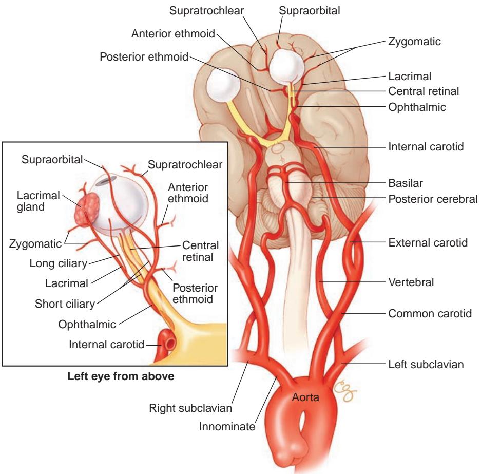
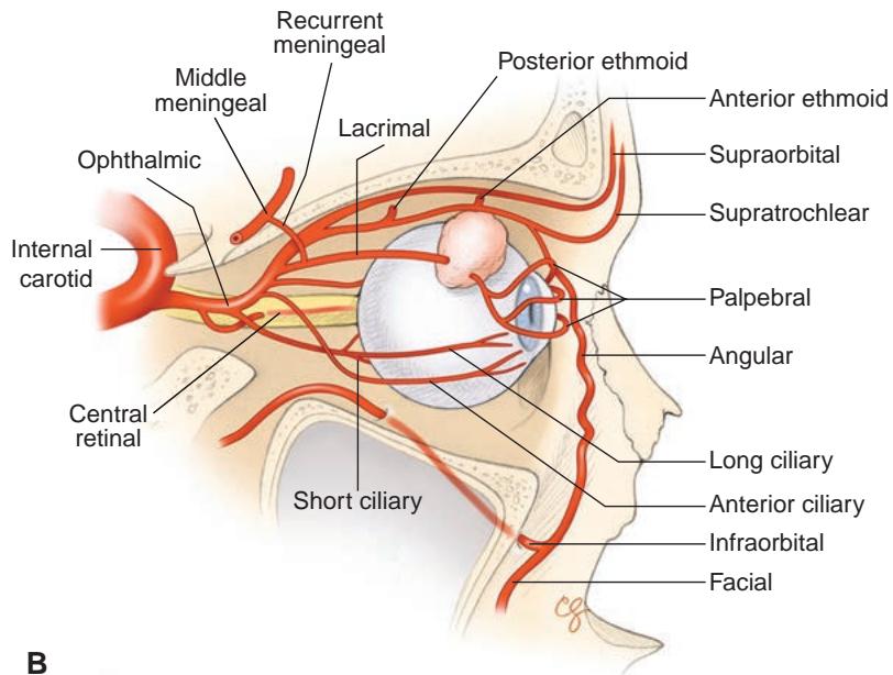
Figure 1-8 Arterial map of the orbit and ocular adnexa. A, Arterial supply of the orbit and ocular adnexa originating from the aortic arch. B, Sagittal view of the arterial supply to the orbit and globe. (Illustrations by Christine Gralapp.)
Anterior Circulation
The common carotid artery divides into external and internal branches at the angle of the jaw (Fig 1-9). The external carotid artery (ECA) supplies blood to the face through its major branches of the facial artery. The scalp is supplied via branches of the superficial tempo ral artery anteriorly and the occipital artery posteriorly. The paranasal sinuses receive their blood supply from branches of the maxillary artery (sphenopalatine and infraorbital), which terminates in the pterygopalatine fossa. The coverings of the brain are supplied by branches of the middle meningeal artery—a major branch of the maxillary artery—which enters the middle cranial fossa through the foramen spinosum, lateral to the foramen ovale. Branches of the middle meningeal artery supply the parasellar area, including the lateral wall of the cavernous sinus, and terminate in the arteries of the foramen rotundum and ovale. Variable meningeal branches may enter the superior orbital fissure. Terminal branches of the facial artery supply the marginal arcades of the eyelids. For further discussion of the blood supply of the eyelids, see BCSC Section 7, Oculofacial Plastic and Orbital Surgery.
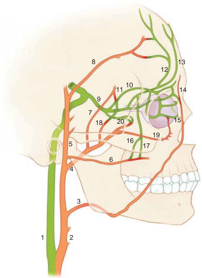
Figure 1-9 Internal carotid artery (green) and external carotid artery (orange) collateral anatomy. 1, internal carotid; 2, external carotid; 3, facial; 4, maxillary; 5, superficial temporal; 6, transverse facial; 7, middle meningeal; 8, frontal branch of superficial temporal; 9, oph thalmic; 10, lacrimal; 11, recurrent meningeal; 12, supraorbital; 13, supratrochlear; 14, angular; 15, palpebral; 16, zygomaticotemporal; 17, zygomaticofacial; 18, deep temporal; 19, infraor bital; 20, muscular. (Modified with permission from Kline LB. Neuro-Ophthalmology Review Manual. 6th ed. Slack; 2008:213. Illustration by Christine Gralapp.)
It is extremely important to understand the extent of the collateral connections be tween the branches of the ECA and the ICA (see Fig 1-9). Interventional neuroradiolo gists and neurosurgeons may inadvertently embolize distal ICA branches, including the central retinal artery (CRA), while placing particles into the ECA. Embolization is most likely to occur during the treatment of arteriovenous malformations, but it can also occur when skull-base tumors are embolized before surgical resection.
The most important collateral connections between the external and internal circula tions traverse the orbit, including the following arteries:
anterior and posterior ethmoidal arteries medially
facial artery where it joins the distal branches of the supratrochlear and infratroch lear arteries around the angular artery medially
infraorbital and supraorbital arteries (including distal connections to the lacrimal artery) anteriorly
zygomaticotemporal branch laterally
various dural branches that traverse the superior and inferior orbital fissures
In rare instances, the OphA may also arise as a branch of the meningeal system of the ECA.
The ICA carries the major blood supply to the intracranial contents (Fig 1-10). The ICA enters the skull through the carotid canal. Within the petrous bone, the artery is near the middle and inner ear, as well as the intrapetrosal portion of CN VII. As the ICA reaches the parasellar area, it turns superiorly just above the foramen lacerum. It then enters the cavernous sinus, where it first gives off the meningohypophyseal trunk and then turns anteriorly to run horizontally parallel to the body of the sphenoid to give off the in ferolateral trunk, which forms anastomoses with branches of the middle meningeal artery. Branches of the meningohypophyseal trunk supply the dura of the posterior cavernous sinus as well as CNs III, IV, V, and VI entering the cavernous sinus. These branches also variably supply the lateral aspect of the sella turcica, including the pituitary capsule and a large portion of the pituitary gland itself.
At the anterior aspect of the cavernous sinus, the ICA loops to reverse its direction under the anterior clinoid and the optic nerve. This loop passes through 2 dural rings, both close to the anterior clinoid (the terminal portion of the lesser wing of the sphenoid). As the ICA passes through the second ring, it becomes intradural (Fig 1-11). Shortly thereafter, the ICA gives off the OphA, which enters the orbit along with the optic nerve through the optic canal. Determining whether an ICA aneurysm is intradural or extradu ral is critical, as the former carries an inherent risk of life-threatening subarachnoid hem orrhage with rupture, whereas the latter is associated with low morbidity with rupture.
Within the orbit, the OphA (see Fig 1-9) may anastomose with recurrent meningeal branches that enter through the superior orbital fissure. The OphA gives off the CRA, which then enters the substance of the optic nerve approximately 10–12 mm posterior to the globe. Within the eye, the CRA divides into superior and inferior arcades. These
A
B