
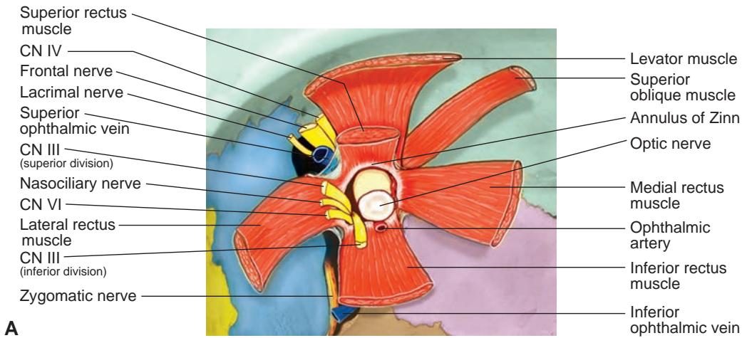
Рисунок 1-5 Анатомия верхушки орбиты. A, 4 прямые мышцы берут начало от кольца Цинна. CN II, III (верхняя и нижняя ветви), VI и носоресничный нерв проходят через кольцо Цинна. CN IV, лобный и слезный нервы, а также глазные вены расположены вне кольца. B, Анатомическая диссекция непосредственно кпереди от верхней глазничной щели. (Иллюстрация части A выполнена Dave Peace; часть B предоставлена Albert L. Rhoton Jr, MD.)
Figure 1-5 Anatomy of the orbital apex. A, The 4 rectus muscles arise from the annulus of Zinn. CNs II, III (superior and inferior branches), and VI and the nasociliary nerve all course through the annulus of Zinn. CN IV, the frontal and lacrimal nerves, and the ophthalmic veins are located outside the annulus. B, Anatomical dissection just anterior to the superior orbital fissure. (Part A illustration by Dave Peace; part B courtesy of Albert L. Rhoton Jr, MD.)
Под верхушкой орбиты в крылонебной области располагаются крылонебный ганглий и верхнечелюстная артерия. Крылонебная область сообщается
Under the apex of the orbit, the pterygomaxillary area contains the sphenopalatine ganglion and the internal maxillary artery. The pterygomaxillary area communicates
спереди через подглазничный канал с щекой и нижним веком
anteriorly through the infraorbital canal to the cheek and lower eyelid
сзади через круглое отверстие и видиев канал со средней черепной ямкой
posteriorly through the foramen rotundum and vidian canal to the middle cranial fossa
сверху через нижнюю глазничную щель с орбитой
superiorly through the inferior orbital fissure to the orbit
B

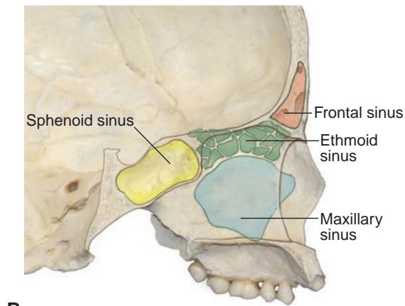
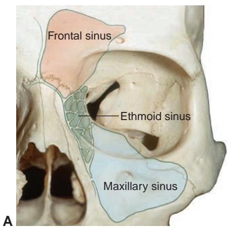
Рисунок 1-6 Анатомическое взаимоотношение 4 околоносовых пазух с орбитой. A, Корональный вид. B, Сагиттальный вид. C, Аксиальный вид. (Иллюстрации выполнены Dave Peace.)
Figure 1-6 Anatomical relationship of the 4 paranasal sinuses to the orbit. A, Coronal view. B, Sagittal view. C, Axial view. (Illustrations by Dave Peace.)
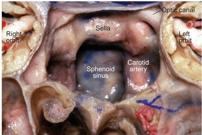
Рисунок 1-7 Корональный срез, вид спереди на клиновидную пазуху, демонстрирующий взаимоотношение внутренней сонной артерии и зрительного нерва в латеральной стенке пазухи. (Предоставлено Albert L. Rhoton Jr, MD.)
Figure 1-7 Coronal section, anterior view into the sphenoid sinus demonstrating the relation ship of the internal carotid artery and optic nerve within the lateral wall of the sinus. (Courtesy of Albert L. Rhoton Jr, MD.)
Каналы и щели
Canals and fissures
Орбита сообщается с окружающими областями через несколько костных каналов и щелей. Сзади орбита граничит с cavernous sinus через верхнюю глазничную щель (см. рис. 1-3). Медиальная стенка орбиты продолжается как латеральная стенка клиновидной кости, отмечая медиальную границу cavernous sinus. При проникновении острых предметов в медиальную часть орбиты они направляются через верхнюю глазничную щель, где могут повредить сонную артерию. В cavernous sinus ятрогенное или травматическое повреждение внутренней сонной артерии (ICA) может привести к формированию прямого высокопоточного каротидно-кавернозного соустья (см. главу 8).
The orbit communicates with the surrounding areas through several bony canals and fis sures. Posteriorly, the orbit is contiguous with the cavernous sinus through the superior orbital fissure (see Fig 1-3). The medial wall of the orbit continues as the lateral wall of the sphenoid bone, marking the medial extent of the cavernous sinus. When sharp objects enter the me dial orbit, they are directed through the superior orbital fissure, where they can lacerate the carotid artery. Within the cavernous sinus, iatrogenic or traumatic damage to the internal ca rotid artery (ICA) may produce a direct, high-flow carotid-cavernous fistula (see Chapter 8).
Орбита соединяется сверху и сзади с передней черепной ямкой посредством зрительного канала (см. рис. 1-2), через который проходят зрительный нерв, OphA и некоторые окулосимпатические волокна (терминальные ветви каротидного сплетения). Снизу, в области верхушки, орбита соединяется с крылонебной ямкой — а следовательно, с височной и нижневисочной областями — через нижнюю глазничную щель. Эта щель, образованная большой крылом клиновидной кости, а также верхнечелюстной, скуловой и небной костями, содержит следующие структуры:
The orbit is connected superiorly and posteriorly to the anterior cranial fossa by way of the optic canal (see Fig 1-2), which transmits the optic nerve, OphA, and some of the oculo sympathetic fibers (terminal branches of the carotid plexus). Inferiorly at the apex, the orbit is connected to the pterygopalatine fossa—and thus the temporal and inferotemporal regions— through the inferior orbital fissure. This fissure, formed by the greater wing of the sphenoid bone and the maxillary, zygomatic, and palatine bones, carries the following structures:
верхнечелюстную ветвь V ЧН (CN V2)
maxillary branch of CN V (CN V2)
подглазничные сосуды
infraorbital vessels
нижнюю глазную вену
inferior ophthalmic vein
ветви крылонебного ганглия
branches from the pterygopalatine ganglion
парасимпатические волокна, иннервирующие слезную железу
parasympathetic fibers that innervate the lacrimal gland
коллатеральные менингеальные артерии, соединяющие наружный и внутренний каротидные круги кровообращения
collateral meningeal arteries that connect the external and internal carotid circulation
Спереди орбита соединяется с нижним носовым ходом (под нижней носовой раковиной) через носослезный проток, по которому слезы поступают в носовую полость. Другие каналы, окружающие орбиту, включают переднее и заднее решетчатые отверстия, через которые проходят кровеносные сосуды, соединяющие внутренний каротидный круг кровообращения (т. е. OphA) с наружным каротидным кругом (т. е. терминальными ветвями решетчатых артерий). Дополнительные надглазничные и скуловисочные отверстия проводят кровеносные сосуды между орбитой и соответствующими сосудами лба и виска.
Anteriorly, the orbit connects to the inferior meatus of the nose (beneath the inferior turbinate) through the nasolacrimal duct, which carries tears into the nasal cavity. Other canals surrounding the orbit include the anterior and posterior ethmoidal foramina, which carry blood vessels that connect the internal carotid circulation (ie, the OphA) to the ex ternal carotid circulation (ie, the terminal branches of the ethmoidal arteries). Additional supraorbital and zygomaticotemporal foramina carry blood vessels between the orbit and corresponding vessels of the forehead and temple.
Rhoton AL, Natori Y. The Orbit and Sellar Region: Microsurgical Anatomy and Operative Approaches. Thieme; 1996.
Rhoton AL, Natori Y. The Orbit and Sellar Region: Microsurgical Anatomy and Operative Approaches. Thieme; 1996.