CATARACT
M. RABINOVICH
The author of this monograph, Mikhail Rabinovich, Doctor of Medical Sciences, is an experienced ophthalmic surgeon. In this work he presents the results of his studies of the problem of the cataract, to which he devoted fifty years of his life.
The monograph begins with a detailed description of a healthy crystalline lens and proceeds to a discussion on the morphology, genesis, and actiology of the different types of cataract, drawing attention to those most frequently encountered. Methods of cataract diagnosis and the indications and contraindications for the various methods of treatment are also dealt with.
(Continued on the back flap)
Much attention is given to the prevention and to the non-surgical and, in particular, surgical treatment of cataracts. Preoperative and postoperative management is discussed in detail. The concluding chapter dwells on the operative results, methods for the correction of postoperative aphakia, and on the dynamics of the improvement of visual acuity occurring with time.
The book is intended for ophthalmologists, medical students, and for all those interested in the problem of the cataract.
BOOKS ON MEDICINE ANT BIOLOGY
ATHEROSCLEROSIS AND THROMBOSIS
MYASNIKOV, A. (Ed.)
This collection is devoted to one of the most urgent problems of cardiology—atherosclerosis and thrombosis of the cardiovascular system.
The articles are concerned with some new aspects of the pathological anatomy of coronary atherosclerosis, pathophysiologic and physiologic mechanisms of the coronary circulation, the pathophysiologic and biochemical changes responsible for disturbances of blood coagulation in coronary atherosclerosis, and clinical observations on biochemical reactions in coronary insufficiency.
Much space is also given to new methods of treatment of coronary atherosclerosis and thrombosis.
It was edited by the late Prof. A. Myasnikov, Fellow of the USSR Academy of Medical Sciences, former Director of the Institute of Therapy, winner of the “Golden Stethoscope” international prize.
The book is intended for therapeutists, pathophysiologists and biochemists.
RICKETS
SVYATKINA, K., KHVUL, A., RASSOLOVA, M.
This monograph is written by a group of eminent specialists in paediatrics.
Their work is based on the latest developments in the study of the aetiology and pathogenesis of rickets. The classification and clinical aspects of rickets are described, much attention is given to the important practical problems of diagnosis and differential diagnosis, prevention, and treatment. Chapters are devoted to hypervitaminosis D, ricketslike osteopathy, and to spasmophilia. Appendices contain a set of physical exercises and massage recommended for infants between six weeks and twelve months old.
The monograph is intended for paediatricians and medical students.
MIR PUBLISHERS
M. PABMHOBAY
RATAPAKTA
H38QATEJIBCTBO “MEQMUMHA*® MOCHBA
M. RABINOVICH
CATARACT
Translated from the Russian
by
Ludmila AKSENOVA, M.D.
MIR PUBLISHERS e MOSCOW
1968
UDC 017.741-004.1=20
REVISED FROM THE 1965 RUSSIAN EDITION
Ha av2autickom sA361Ke
CONTENTS
Preface.. . . . . 2. . 6
CHAPTER 1. Clinical Anatomy and Physiology of the Crystalline Lens
Cataract-Pathological Conditionof the Lens . 14
Diagnosis 35
Indications and Contraindications for Treatment 41
Prevention and Non-surgical Treatment oe . . D3
Surgical Treatment 63
Preoperative Management 65
Operation . 74
Postoperative Period 124
Complications 127
Postoperative Results. Aphakia . 153
Bibliography . 162
Index of Names 171
Subject Index . 173
PREFACE
In this work we present the results of our fifty-year’s experience in ophthalmology devoted to that vitally important problem in this branch of medicine, cataract.
The subject of cataract which embraces studies dealing with its pathogenesis, prophylaxis, and treatment is still a most urgent and vitally important problem of ophthalmology.
The practicing ophthalmologist is mainly interested in the methods of prevention and treatment, and for that reason the greater part of this work is concerned with that aspect of the problem. Prevention is a science which is only just beginning or may, perhaps, be said to have just begun at any rate, even though in its infancy it has a promising future. As to the treatment (to be more precise, treatment of the patient suffering from cataract) it may be divided into non-surgical and surgical. We use the term “non-surgical” to designate all types of active and advanced therapy, with the exception of the surgery, instead of the rather archaic term “conservative treatment”.
M. Rabinovich, D.Sc. (Med.)
CHAPTER 1

CLINICAL ANATOMY AND PHYSIOLOGY OF THE CRYSTALLINE LENS
The crystalline lens (lens crystallina), an important constituent of the optical system of the eyeball, is a transparent, semi-solid, biconvex body. It has anterior and posterior surfaces with centres known as poles; the wavy line dividing these surfaces from one another is called the equator and lies a little behind of the ciliary processes. The equatorial diameter of the lens is nine to ten millimetres, its antero-posterior axis connecting the anterior and posterior poles is about 3.6 millimetres. The crystalline lens lies between the iris and the vitreous body, lodged in a saucer-like depression on the anterior surface of the latter (fossa patellaris). The lens and the vitreous body, however, are not in direct contact, being divided by a retrolental capillary space (spatium retrolentale). The periphery of the iris is separated from the anterior surface of the lens by the aqueous humour of the posterior chamber, while its pupillary zone corresponds to the central part of this surface. During dilatation or contraction of the pupil the iris slides over the anterior surface of the lens. This surface is less convex than the posterior: its radius of curvature is ten millimetres, while that of the posterior is six millimetres (Fig. 1).
The accommodative function of the lens effected by its alternating thickening and flattening would not be possible, were it closely joined to the surrounding internal structures of the eyeball.
The crystalline lens consists of two main structures: a non-cellular hyaline capsule, and the lens proper formed of epithelium and fibres. Anterior and posterior parts are distinguished in the capsule. The anterior capsule (between 0.008 and 0.02 millimetres thick) thickens with age. Its external layers facing the iris at the equator constitute the zonular lamella, formed of fibres of the ciliary zonule (zonule of Zinn) interwoven with the capsule, and are readily separated from the capsule itself. The posterior capsule is much thinner, its thickness at the posterior pole being between 0.002 and 0.004 millimetres.
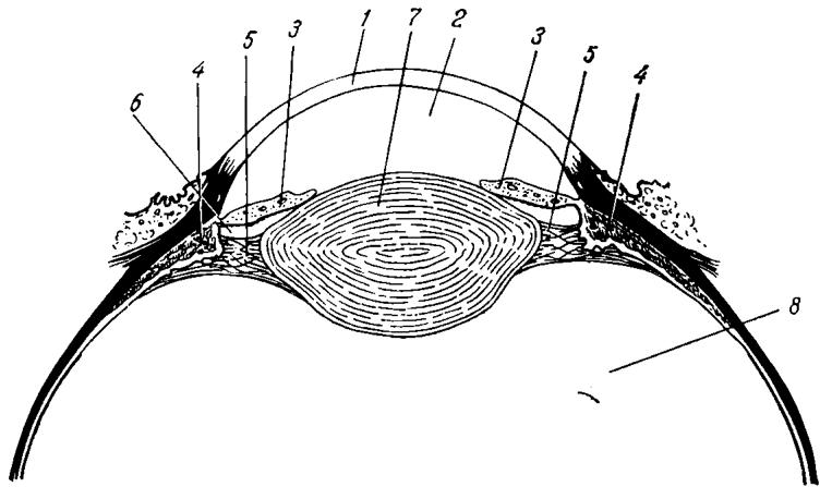
Fig. 1 Anterior segment of the eyeball
1 — cornea; 2 — anterior chamber; 3 — iris; ¢— ciliary body; 5 — fibresof the ciliary zonule; 6 — posterior chamber; 7 — crystalline lens; 8 — vitreous body
The internal layer of the anterior capsule is a product of the epithelial cells which line the back of this capsule (the posterior capsule has no epithelium). Nearer to the equator the epithelial cells are arranged in regular meridional rows, which, gradually becoming longer, are transformed into the lens fibres. Electron microscopy (Cohen, 1965) has shown that the latter arc furnished with dents by which they become firmly joined during accommodation. The fibres are shaped like hexagonal prisms. The growing fibre-cells in each meridionally arranged row push the older fibres to the centre of the lens, causing them to be superimposed on one another like the segments of an orange and thus forming radial lamellae. Adjoining lamellae form layers. The fibres grow toward the poles, but never reach them extending only to the so-called lens star. The rays of the star (nine or twelve in number) are formed by radial lines, and divide the surface of the lens into sectors. New fibres develop throughout life. It might be expected that the lens, enclosed in a dense capsule, would grow continuously in volume, but this does not happen. The lens loses water with age, undergoes sclerosis, and becomes thickened at the centre. This thickening begins as early as 25 or 30 years of age, in an individual over 40 the lens centre becomes a dense nucleus, while its peripheral parts, the cortical layer, remain relatively soft. The transition from soft cortex to dense nucleus occurs gradually. Owing to this process the volume of the lens remains relatively constant at all stages of life.
Lens structure has been studied’in detail by means of biomicroscopy. Its substance is not homogeneous in density and because of this, so-called zones of discontinuity are seen in it on examination by slit lamp. The lens centre is poorly illuminated; it is bounded by two bright zones which together have the shape of a bean and are known as the central bands of the foetal nucleus. The peripheral bands of the nucleus circumscribe the central ones and are not as bright. They appear by the time of birth. More peripherally are two bright bands, of the central nucleus; like the lens star, they appear about the age of ten and become more pronounced with years. Zones of discontinuity are seen nearer to the periphery, they become dim with age and serve as orienting points for locating the site of opacity.
The lens is held in place by thin threads of the ciliary zonule (see Fig. 1) which consists of many smooth, elastic, structureless fibres originating in the epithelium of the basal lamina of the ciliary body and in the depression between the ciliary processes. The ciliary zonule has a peculiar structure, the fibres originating on its front surface being interwoven into the region of the equator and the posterior capsule of the lens, and those originating on its back surface, into the anterior capsule. By this structure a space is formed between the intertwining fibres, known as Petit’s canal, the zonular space, or the spatia zonularia. The ciliary zonule is quite strong in young individuals; with advanced age, particularly in old people, it becomes fragile and tears easily. This fact is of essential importance in surgical removal of a cataract.
Chemical composition of the lens. About 65 per cent of the total weight of the lens is water. During growth this amount increases to 75 per cent but then decreases greatly with age, the lens undergoing dehydration. About 65 or 75 per cent of the total water content is confined to the elements of the cortical layer, since it is formed from the youngest fibres. The greater part of the water occurs in a free state; a small part is bound.
Proteins make up about 35 per cent of the mass of the lens. It may be noted here that the lens, particularly its nucleus, contains more protein than any other human tissue. Both soluble and insoluble proteins are present. In young individuals the proteins are almost all soluble; they decrease, however, with age, while the amount of insoluble proteins (albuminoids) increases.
The amount of cysteine (an organic compound belonging to the group of amino acids) in the insoluble proteins is negligible, but is quite large in the soluble ones. Cysteine plays a very important role in the vital activity of the lens, taking part in the oxidation processes occurring within it (internal respiration). Its content also decreases in advanced age and tissue respiration in the lens consequently diminishes.
In addition to these constituents, the crystalline lens contains a small amount of lipoids, cholesterol among them, about 2 per cent of inorganic compounds (calcium, phosphorus, etc.), and some ascorbic acid, its amount decreasing with age.
Finally, mention must be made of one further component of the lens tissue, glutathione, which also plays an important role in tissue respiration.
The functions performed by the lens do not require high metabolic activity. Energy is needed only to maintain transparency and to provide the nourishment necessary for the formation and growth of new fibres, and to preserve the elasticity of the capsule. The lens has no system of blood vessels and is supplied with nutrients by mutually supported metabolic processes occurring within it and the aqueous humour. Because of this, qualitative and quantitative changes taking place in the aqueous humour may affect the condition of the lens in one way or another.
From the point of view of colloidal chemistry, the lens is a complex system which maintains its transparency only under certain conditions: it becomes opaque both with the loss of water, and with an increase in its content, with an increase in the concentration of various substances in the anterior chamber, etc. Its optical properties also depend on the permeability of the anterior and posterior capsules.
The lens is a system in a state of an unstable equilibrium that can only be maintained by a continuous supply of energy. In Fradkin’s apt expression (1947), we would do better to think about the factors facilitating maintenance of transparency rather than about the causes of opacification. From this point of view, the fact that the loss of transparency and opacity of the lens are a relatively frequent occurrence should cause no surprise.
Дальше — с подпиской Awamed+
Осталось ещё 168 страниц источника. Подписка открывает материал целиком вместе с оригиналом, рисунками и файлами.